|
Return
to |
|
Crime Scene Investigation (CSI) with Powdery Mildew Fungi.This exercise can be used to stimulate the investigative nature of your students as they use forensic plant pathology techniques with powdery mildew cleistothecia (the sexual stage of powdery mildew fungi) to prove their innocence in a mock murder investigation. The exercise is suitable for grades 7 -12 and does not require the purchase or maintenance of special cultures, since cleistothecia produced by powdery mildew fungi are easy to find on plants in nature. Dissecting and compound microscopes are required. Ruhl, G.. 2004. Crime Scene Investigation (CSI) with Powdery Mildew Fungi. The Plant Health Instructor. DOI: 10.1094/PHI-K-2004-0330-02
Copyright
© 2004 by The American Phytopathological Society |

|
|
|
Crime Scene Investigation (CSI) with Powdery Mildew FungiPURPOSE AND BACKGROUND INFORMATIONPurpose:This exercise can be used to stimulate the investigative nature of your students as they use forensic plant pathology techniques with powdery mildew cleistothecia (the sexual stage of powdery mildew fungi) to prove their innocence in a mock murder investigation. In order for students to truly understand the nature of science and technology, they must model the process of scientific investigation through inquiries. This lesson is suitable for grades 7 -12 and does not require the purchase or maintenance of special cultures, since cleistothecia produced by powdery mildew fungi are easy to find on plants in nature. Teachers will use leaves infected with powdery mildew fungi that they have previously collected. Note: This lab works best in regions that experience cold weather for part of the year, since colder temperatures at the end of the plant's growing season encourages the production of cleistothecia (sexual stage of the powdery mildew fungi) on leaves. This lab may not work as well in areas where warm weather prevails year-round, since this type of weather promotes primarily the conidial (asexual) stage of powdery mildew fungi, with sparce to no cleistothecia production. Background information:CRIME SCENARIO: An eyewitness thought he saw you and two others running from a wooded park where a murder took place. The police have brought you all in for questioning and during their investigation they notice that the leaf pieces stuck to all of your shoelaces have the same powdery-like appearance as the leaf pieces that were found on the murder victim's clothes. Although only circumstantial, the public is demanding the arrest of a suspect, and it appears that you are going to be arrested for murder unless you can prove them wrong. You must delve into the depths of forensic plant pathology and use diagnostic skills and tools to prove your innocence…that is, if you ARE innocent! FORENSIC BACKGROUND INFORMATION:
Additional background information and color images are available in the K-12 Lab: Powdery Mildew Fungi: Classification and Ecology. Disclaimer: Powdery mildew fungi have long been classified to genus based on the number of asci contained in the cleistothecium and on the morphology of the hyphal appendages growing out of the wall of the cleistothecium. Recently, new research has resulted in the reclassification of many of these genera according to the phylogeny (an estimate of evolutionary relationships) inferred from the DNA sequence of the ribosomal ITS region and a number of morphological characteristics. In particular, the morphology of the conidia (asexual spores) and conidiophores (the structures that produce the conidia) is important in the new classifications. Despite these changes, this lab can still be used to teach students about forensic evidence, the use of identification keys, and morphological variation among fungal species. Copyright
© 2004 by The American Phytopathological Society |
||||||


|
Return to Powdery |
|
Powdery Mildew Fungi: Classification and Ecology Purpose and Background Information Purpose: Powdery mildew fungi are ideal for use in a laboratory exercise on fungal classification, fungal spore types, host-parasite interaction during an ecology unit, or a study of host range. Students will discover how many different plant hosts they can find that are infected by the same genus of a powdery mildew fungus, or how many different genera of powdery mildew fungi can be found on the same plant host. This exercise demonstrates the diversity that exists within a fungal order. With a good collection of leaves infected with different powdery mildew fungi, students learn to use a written key and/or an illustrated key (or could even make their own key) to identify the powdery mildew fungus to genus. Since powdery mildew fungi reproduce by means of two spore types: asexual spores (conidia) and sexual spores (ascospores), discussions of the types of reproduction in fungi would be facilitated. Background information: Powdery mildews are one of the most common, conspicuous, widespread, and easily recognizable plant diseases. As a group, powdery mildew fungi infect many species of plants, including many trees and shrubs, numerous ornamentals, vegetables, cereals, grasses, and even weeds. However, individual species of powdery mildew fungi are usually very host specific. For example, the species of fungus causing powdery mildew on pumpkin is different from that causing the disease on roses. Extensive losses in plant growth and crop yield occur annually due to powdery mildew. The primary sign of powdery mildew is grayish white, powdery blotches on leaves. Usually, powdery fungal growth appears first on the upper leaf surface (FIGURE 1). Eventually the entire leaf may become covered with mildew (FIGURE 2). Powdery mildew fungi also can infect other parts of the plant and may cause distortion and stunting of shoots, leaves (FIGURE 3), and flowers (FIGURE 4), and russeting on fruit (FIGURE 5). The type and extent of symptoms vary depending on the combination of powdery mildew fungal species and host plant species involved. Late in summer and fall, the sexual stage of many species of powdery mildew fungi, the cleistothecia, is visible as black or brown, pinhead-sized, spherical specks among the white to grayish mildew mycelium in the older infected areas on the leaves of many plants (FIGURE 6). (These are the leaves that should be collected and pressed flat between newspaper for storage and use in this laboratory exercise.)
The powdery mildew diseases of various crops and other plants are caused by many different species of fungi grouped into six main genera in the order Erysiphales, an order that includes a single family, the Erysiphaceae. The fungi causing powdery mildews are obligate parasites (or biotrophs), meaning that they cannot be cultured on nutrient media and require a living host to grow. These fungi reproduce by means of two spore types: asexual spores called conidia and sexual spores called ascospores. The conidia are usually barrel-shaped or oval and are usually formed in chains (FIGURE 7) at the ends of specialized hyphae called conidiophores produced from the mycelium growing on the surface of a plant’s leaves, stems, flowers or buds. The combination of the mycelium, conidia and conidiophores gives the leaf surface a powdery appearance from which the name powdery mildew is derived. The fungi are spread when the conidia are carried by air currents to new plant surfaces. Under favorable conditions, the fungal spores germinate (FIGURE 8). Unlike most fungal parasites that invade plant tissues, most powdery mildew fungi grow superficially on top of the leaf surface (FIGURE 9). Fine, thread-like infection pegs penetrate the epidermal cells of the leaves and form haustoria (FIGURE 10). Haustoria are specialized hyphae for nutrient absorption from the plant cells. The plant is damaged by the loss of nutrients to the fungus, disruption of photosynthesis, and premature death of leaves or other infected plant tissues.
When environmental conditions or nutrition become unfavorable for growth (usually later in the growing season), the fungus shifts to the sexual stage and produces cleistothecia (sing. cleistothecium). The cleistothecia are closed, thick-walled, tiny, black, spherical structures (white to tan when young) that house sacs called asci (sing. ascus) (FIGURE 11 and FIGURE 12). The oval sexual spores, called ascospores, are produced within the asci. Fungi that produce ascospores in asci belong to the Ascomycetes, a fungal group that includes yeast, morels, and many plant parasites.
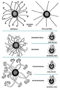
Cleistothecia have ‘arm-like’ appendages that radiate out from their outer surface. Inside each cleistothecium is a single ascus or many asci. Currently, the powdery mildew fungi are classified to genus based on the number of asci contained in the cleistothecium and on the morphology (physical appearance) of the hyphal appendages (arms) growing out of the wall of the cleistothecium (FIGURE 11, FIGURE 12, and Key to Genera of Powdery Mildew). Recently, Saenz and Taylor (1999) have proposed that powdery mildew genera be reclassified according to the phylogeny (an estimate of evolutionary relationships) inferred from the DNA sequence of the ribosomal ITS region and a number of morphological characteristics. If this occurs, the morphology of conidia and conidiophores will be used as the primary characters for classification. Copyright © 2004 |


|
|
|
Crime Scene Investigation (CSI) with Powdery Mildew FungiMATERIALS AND METHODSCollection Methods:In late summer or autumn collect leaves with mature (black) cleistothecia on plants that show signs (pale, dusty-white coating on leaves) of powdery mildew. The cleistothecia should be visible to your eye without the aid of a magnifying glass. Asters, azalea, cherry, filbert, grape, horse chestnut, lilac, oak, phlox, rose, zinnia, evening primrose (Oenothera spp., often called sundrops), viburnum, wheat, willow and many other shrubs, trees, flowers, weeds and garden plants are good sources of cleistothecia. Leaves exhibiting abundant cleistothecia are good specimens for teaching purposes. Infected leaves should be pressed flat between pieces of newspaper. Pressed, dried, leaf specimens can be stored in envelopes for use over many years. Label the envelope with the pressed leaves giving the name of the plant, the powdery mildew genus (you will need to identify the genus with the forensic key and diagrams), and location of collection, for future reference. For your own purposes of cataloguing and ease of choosing teaching material for labs, you may want to number your envelopes and keep a separate list of the specific genera of powdery mildew found to be associated with each plant collected. You will find that several different genera of powdery mildew will infect the same host type. For example, Microsphaera and Phyllactinia may both be found on oak. This is why it is important to label your envelopes by powdery mildew genus and not host plant. Lab Preparation:
Lab Set-up:
Procedures for students:FORENSIC TOOLS:You are a suspect in a murder investigation. You must attempt to prove your innocence and clear your name using dissecting and compound microscopes, clear tape, petri dishes, forceps, microscope slides, a water dropper and a forensic key with the special characteristics of powdery mildew cleistothecia appendages. FORENSIC LAB PROCEDURES:You may work alone or in a group of three or four.
Repeat Sections B and C (Examination and Identification of Evidence) with the plant material in your petri dish labeled "Murder Victim" including the recording of results. Forensic Key and Diagrams to the Genera of Powdery Mildew FungiAppendages coiled or hooked at tip - Uncinula
Locate the Forensic Evidence Data Table on the Case Study Sheet . Record and draw the type of appendages (arms) found on the surface of the cleistothecia (e.g. hooked tip; bulb-like base; antler-like dichotomous branching; or simple, string-like arms), the number of asci (one or several) contained within each cleistothecium, and the name of the powdery mildew genus.
Copyright
© 2004 by The American Phytopathological Society |
|||||||||||||||||||||||||||


|
|
|
Crime Scene Investigation (CSI) with Powdery Mildew FungiLESSON PLANLearning objectives:To learn about a specific method of identifying fungi, using a written key and an illustrated key. Powdery mildew fungi can be identified to genus by the morphology (appearance) of the sexual stage (cleistothecia). To learn about scientific investigation using forensic techniques. Exercise description:Plant leaves infected with powdery mildew disease are collected after the sexual stage, the cleistothecia, are visible on the leaf surface. Cleistothecia from leaf material are examined with a compound microscope to identify the type of appendages present. The fungi are identified to genus using the appendage type and the number of asci (spore-containing sacs) inside each cleistothecium using both a written and an illustrated key. The students, using forensic plant pathology techniques, must attempt to prove themselves innocent in this CSI lab. Images for class use are in the Supplementary Information and References section. Time frame:Infected leaves can be collected in late summer or autumn, pressed flat, and dried to preserve them until used. Fresh leaves also may be examined. Once you have a collection of powdery mildew infected leaves, the actual exercise takes little preparation and can be done at any time. Study questions:Forensic Conclusions: Innocent or Guilty?
Copyright
© 2004 by The American Phytopathological Society |
|
|
|
Crime Scene Investigation (CSI) with Powdery Mildew FungiSUPPLEMENTARY INFORMATION AND REFERENCESSupplementary Information:Websites: “Identification of Powdery Mildew Fungi” by V. Heffer,
M. L. Powelson, and K. B. Johnson, APSnet Feature “Control of Powdery Mildew
Using the UC Davis Powdery Mildew Risk Index” by W. D. Gubler,
M. R. Rademacher, S. J. Vasquez, and C. S. Thomas. References:Agrios, G. N. 1997. Plant Pathology, 4th ed.
“The Powdery Mildews,” pp.295-298. Academic Press, Carroll, J. E. 1994. Learning Biology with Plant Pathology. Nameth, S. and J. Chatfield. Powdery mildews on
ornamental plants. Pecknold. P. C. Powdery Mildews of Ornamentals. Ruhl, G.E. and Saenz, G. S. and J.W. Taylor. 1999. Phylogeny of the Erysiphales (powdery mildews) inferred from internal transcribed spacer ribosomal DNA sequences. Canadian Journal of Botany 77:150-168. Weber, R. W. S. and J. Webster. 2001. Teaching techniques for mycology: 13. Functioning of cleistothecia in Phyllactinia gutatta. Mycologist 15:26-30. Webster, J. 1980. Introduction to Fungi, 2nd ed. Yarwood, C. E. 1973. Pyrenomycetes:
Erysiphales. Pages 71-86 in: The Fungi, An Advanced
Treatise. Volume IVA. G.C. Ainsworth, F.K. Sparrow, and A.S. Sussman, A. S., eds. Academic Press, Yarwood, C. E. 1978. History and taxonomy of the
powdery mildews. Pages 1-37 in The Powdery Mildew. D.M. Spencer, ed. Academic
Press, Copyright
© 2004 by The American Phytopathological Society |
|
|
|
Crime Scene Investigation (CSI) with Powdery Mildew FungiMATERIALS FOR CLASS USEForensic Key and Diagrams to the Genera of Powdery Mildew Fungi Copyright
© 2004 by The American Phytopathological Society |

Forensic Key and Diagrams to the
Genera of Powdery Mildew Fungi
Appendages coiled or hooked at tip - Uncinula
Appendages simple and straight with bulb-like base - Phyllactinia
Appendages branching dichotomously (antler-like at tip)
Cleistothecium contains a
single ascus - Podosphaera
Cleistothecium contains
several asci - Microsphaera
Appendages simple or irregularly branched, often entangled
Cleistothecium contains a
single ascus - Sphaerotheca
Cleistothecium contains
several asci - Erysiphe
Key to Genera of Powdery
Mildew
(Drawing courtesy of C.B. Kenaga, E.B. Williams, and
R.J. Green)
CASE STUDY SHEETInvestigators: _________________ CHAIN OF EVIDENCE TABLE
Forensic Conclusions: Innocent or Guilty?1) Compare the forensic plant pathology observations you have recorded for the two different samples. Are the appendages observed on the two different samples the same or are they different?
2) If the appendages you observed on the cleistothecia found on the plant material taken from your shoelaces (murder suspect) are different from appendages on the cleistothecia found on the plant material taken from the murder victim, what does this evidence suggest?
3) If the appendages on the cleistothecia found on the plant material taken from your shoelaces (murder suspect) are the same as the appendages on the cleistothecia found on the plant material taken from the murder victim what does this evidence suggest?
4) Do you need an alibi as to where you were when the murder took place? Explain.
|
|||||||||||||||||||||||||||||||||||||||||||||||||||||||||||||||||||||