بسم الله الرحمن الرحيم

In the name of Allah, Most Gracious, Most Merciful

دكتور محمد محمد الحناوى

أخصائى النساء والتوليد والعقم والنساء الأصحاء - خبير تنظيم الأسرة -عضو الجمعية المصرية للخصوبة والعقم - الكشف بالأشعة التليفزيونية - جمهورية مصر العربية- محاقظة دمياط- مدينة رأس البر- ت 529416 محمول 0122503011

 الصفحة الرئيسية       من أنا      عيادتى        عائلتى      أتصل بى       وطنى     الصور      مواقع طبية      زملائى      الدليل الطبى


colposcopy

 The colposcope
is a binocular
 low power  microscope,which helps the gynaecologist to perform

 colposcopy. because It is impossible to diagnose diseases or other problems simply by looking at the

 cervix with the naked eye. A magnified view is necessary to find any abnormalities

A colposcopy is done by a specially trained doctor  (colposcopist) in a clinic or  outpatient

 The examination takes 10 - 20 minutes but the whole appointment may take about an hour

This exam usually is done between menstrual periods

patient must not use anything in the vagina for 24-48 hours before the procedure --includes

 spermicides, vaginal medications, douching products or tampons or vaginal sex  because These all

 interfere with the accuracy of the test

The colposcope enables the gynaecologist to look at the cervix ,vagina and

 external genital area in detail

 with magnification (The Colposcope is not inserted into the vagina.),

in particular the area in cervix where the smear was taken from (usually

 the transformation zone).


Methods of Colposcopic Examination
Classical or Extended Colposcopy
The cervix and vagina are first examined at magnification of 7x or 10x following which excess mucus is removed from the cervix
Acetic acid 3% to 5% is applied by cotton swab.Abnormal epithelium appear as thick white (acteo-white)
Schiller iodine test may be applied

The doctor will use a speculum,The Pap Smear is done as usual ,

under 2 - 3 different magnifications,

Any excess mucus or other secretions will be cleaned (The Saline Technique)

from the cervix using a large cotton swab ,

the application of acetic acid, a vinegar solution (dissolves mucous and accentuates atypical areas

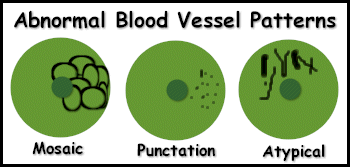
 (white epithelium, punctation, mosaic and atypical vessels) by causing cellular dehydration and
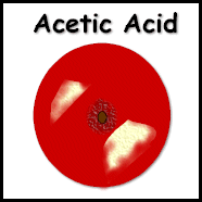
 coagulation of cellular protein--The effect of the acetic acid peaks in

 approximately 2 minutes and

 fades in approximately 5 minutes thus doctor may need to re-apply the

 acetic acid solution severa  timesWhite epithelium is sometimes associated

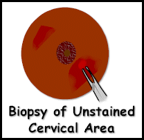
 with dysplasia.  These areas will be biopsied by doctor near the end of

 the procedure ,While the acetic acid is used different colored filters to be able to see the blood

 vessel patterns that can't be seen using regular light. 

 The green filter absorbs  the red color so that the pattern of the cervical blood vessels can be seen


paint the cervix and vagina with an iodine solution called Lugol's Solution.  This stains the glycogen,

a component of cells.  Mature, normal cells will stain a dark-brown color.

  Immature cells, cervicitis, and dyspasia cells will not stain.  This is

 called a Schiller Test.   Non-staining areas will biopsied.

These are noted using the hands of the clock, i.e.. 10:00, 1:00, etc.

A scraping if the endocervical canal (ECC) will be done by a thin instrument

 called an endocervical curette ,

doctor will apply pressure to the cervix using a large cotton swab to stop any bleeding from the biopsy

 areas. 
 
After examining the cervix and deciding what the appearance of the cervix is

 (again this is subjective),

the gynaecologist will make one of three decisions:

               1- The cervix looks entirely normal
 
               2- The cervix appears mildly abnormal   (known as a low grade

 abnormality)

               3- The cervix appears significantly abnormal  (known as a high grade abnormality)


All of these terms Below  are roughly equivalent and are often used interchangeably.

Biopsy Grading Smear Grading Colposcopy Grading
Normal Normal Normal
- Borderline Low Grade
CIN 1 Mild Low Grade
CIN 2 Moderate High Grade
CIN 3 Severe High Grade


Aided Visual Inspection of the Cervix
"Acetic Acid Test"(AAT)

 
Normal cervix, acetic acid pool visible at 6 o'clock. No medical intervention required.
Call for re-screening according to established policy.
Normal cervix, negative Acetic Acid Test (AAT), small condyloma acuminatum on left vaginal wall (9 o'clock). No medical intervention required.
Call for re-screening according to established policy.
Normal cervix, negative AAT. Some degree of ectopy is visible and subsequently, the squamo-columnar junction line is clearly visible. White endocervical mucous is present. No medical intervention required.
Call for re-screening according to established policy.
Severe postmenopausal atrophy of the squamous epithelium, negative AAT. Refer the patient to Primary Health Clinic for treatment if patient symptomatic.
Cervical polyp, negative AAT. No medical intervention required.
Call for re-screening according to established policy.
Normal cervix, negative AAT. Ectopy is present with metaplastic epitheliumgrowing medially at 12 o'clock (containing crypt openings). Posteriorly several "bands" of metaplastic epithelium are visible. Outside the squamo-columnar junction line the transformation zone is visible as a slightly white circular area. No medical intervention required.
Call for re-screening according to established policy.
Marked ectopy, negative AAT. No medical intervention required.
Call for re-screening according to established policy.
Nabothian cyst at 5 o'clock. Atypical acetowhite lesion at 11 o'clock extending up into the canal - colposcopy indicated. Take swab for culture (if facilities available).
Refer the patient to Primary Health Clinic.
Atypical lesion anteriorly, positive AAT - repeat screening in 6 month's time. Refer the patient to Primary Health Clinic.
Acetowhite metaplastic epithelium anterior and posterior. Atypical lesion at 12 o'clock (at the periphery). Refer the patient to Primary Health Clinic.
Condylomata acuminata at 10 o'clock. Refer the patient to Primary Health Clinic.
Negative with acetowhite metaplasia. Crypt openings are present within metaplastic epithelium. At 1 o'clock Nabothian cyst is present (yellow). False negative AAT. Refer the patient to Primary Health Clinic.
Normal with acetowhite metaplasia in the transformation zone. False positive AAT. Refer the patient to Primary Health Clinic.
Condylomata acuminata at 6 o'clock, acetowhite metaplasia anterior. Refer the patient to Primary Health Clinic.
Atypical acetowhite lesion extending up into the canal - colposcopy and biopsy indicated. Refer the patient to Primary Health Clinic.
Positive AAT. Probably normal, but a biopsy is desired in order to rule out cancer. Refer the patient to Primary Health Clinic.
Positive AAT. Probably normal, but abnormal blood vessels indicate biopsy. Refer the patient to Primary Health Clinic.
Low grade SIL (CIN I) at 12 o'clock with acetowhite metaplastic epithelium posterior. Refer the patient to Primary Health Clinic.
Positive AAT, low grade SIL (condylomata acuminata). Refer the patient to Primary Health Clinic.
Positive AAT, high grade SIL anterior (CIN II). Refer the patient to Primary Health Clinic.
Positive AAT, high grade SIL (CIN III) at 5 o'clock. Acetowhite metaplastic epithelium anterior. Refer the patient to Primary Health Clinic.
Leukoplakia before application of acetic acid; probably high grade SIL (CIN III). Refer the patient to Oncology Centre.
Infiltrating cancer. Refer the patient to Oncology Centre.

 الصفحة الرئيسية       من أنا      عيادتى        عائلتى      أتصل بى       وطنى     الصور      مواقع طبية      زملائى      الدليل الطبى