
 HOME
HOME  EMAIL ME
PICTORIAL TOUR OF A LAPAROSCOPIC GALL BLADDER REMOVAL

OVERVIEW
The picture on the left is a representation of the organs surrounding the gall bladder. The liver produces bile which flows down to the intestine in a tube called the "Bile duct". The gall bladder is a bag-like organ which is like a branch on the right side of the bile duct. The gall bladder is stuck to the liver, and is fixed to the bile duct by the "cystic duct" and "cystic artery" - both of which have to be accurately found by the surgeon, clips applied to prevent leakage or bleeding and then cut in order to release the gall bladder before removing it.
Below is a description of steps involved in removing a gall bladder in Laparoscopic Surgery. Clicking on each thumbnail will load an image of one step of an actual laparoscopic operation.
CLICK ON THUMBNAILS TO DISPLAY IMAGE
Picture 1
The outside view of the patient's abdomen shows tubes, called "cannulas" going into the abdominal cavity through punctures. Instruments and the telescope are passed through these cannulas
 Picture 2
The gall bladder is held up to try and get to its main attachments (seen at lower right) from which it must be freed.
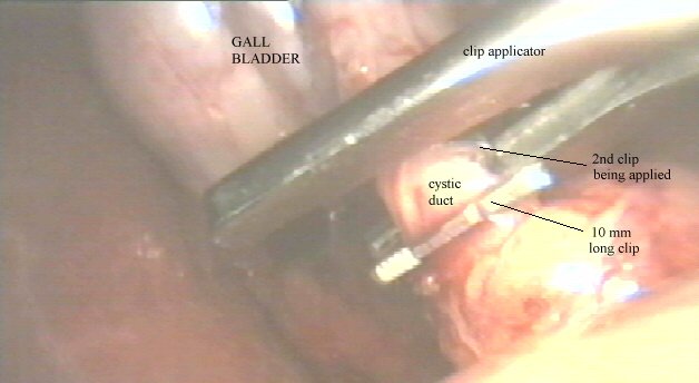 Picture 3
The first important attachment is the "cystic duct" - the narrow tube that connects the gall bladder to the main bile passage - the Bile Duct. Titanium clips are used to clip the cystic duct so as to to prevent leakage of bile from the cut ends after it is cut.The great magnification makes a small clip appear huge.
 Picture 4
The clipped cystic duct is cut with a pair of laparoscopic "hook" scissors.
 Picture 5
The gall bladder is now attached only by the artery. The picture show a blunt instrument that is used to locate the artery before applying clips and cutting it as in the previous pictures.
 Picture 6
This image, blurred by some blood on the lens, shows the completely freed gall bladder lying in a small space between the liver and the stomach wall just before being pulled out.
 Picture 7
This picture shows an inside view of the wall of the abdomen. Half the gall bladder has been pulled out through a puncture and is invisible, the remaining half is seen as if suspended from the abdominal wall above. The emerging outer half of the gall bladder can be seen in the next picture
 Picture 8
In this outside view we see the gall bladder emerging from one of the punctures
 Picture 9
The small size of a gall bladder is highlighted in comparison with a scale
 Picture10
This picture shows the minute scars left after a laparoscopic operation - in this instance - an appendix removal
HOME EMAIL ME