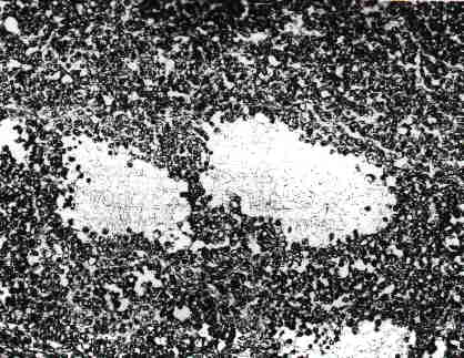
lmmunohistochemistry for p53 Protein
Small Cell Carcinoma of the Ovary of the Hypercalcemic Type: p53 Protein Accumulation and Clinicopathologic Features
JEFFREY D. SEIDMAN, M.D.
Twenty women with ovarian small cell carcinoma of the hypercalcemic type were studied. Mean patient age was 24 years, 11 had hypercalcemia, and the 2-year actuarial survival was 18%; there was only one long-term survivor. Twelve of 15 tumors tested (80%) exhibited p53 protein accumulation by immunohistochemistry, supporting the presence of p53 gene mutations in these tumors. p53 gene mutations may have an important role in the evolution of small cell ovarian carcinomas. ~ 1995 Academic Press, Inc.
The p53 gene is a tumor suppressor gene located on the short arm of chromosome 17 and encodes a 53-kDa nuclear phosphoprotein that is found at low levels in virtually all cells. The p53 protein binds to specific regions of DNA and is believed to be a negative regulator of the cell cycle. MutaŁtion of the p53 gene is the most common genetic event described in human malignancies [1, 2]. A substantial proportion of ovarian carcinomas contain p53 gene mutations [3-11] and/or accumulate p53 protein [12-16].
Although a rare tumor, small cell carcinoma (hypercalcemic type) of the ovary is of special importance for two reasons. First. it occurs in young women with a mean age of 23 years, and second, it is one of the most rapidly fatal ovarian tumors [17-28]. Despite the extensive literature on p53 mutation in ovarian carcinomas, there is little informaŁtion available on the status of p53 in this tumor. Small cell carcinoma has clinical and pathologic features that are markŁedly different from the majority of ovarian carcinomas, and therefore, information on p53 in ovarian carcinomas may not be applicable to this particular tumor type. We performed immunohistochemical analysis on a series of ovarian small cell carcinomas using an antibody to the p53 protein to eluciŁdate the status of the p53 gene in these tumors.
A series of small cell carcinomas were retrieved from the AFIP files for the period 1980-1993. Cases were included in the study if they fit the description of small cell carcinoma of the ovary {hypercalcemic type) as previously described [17, 29, 30].
Clinical information was tabulated from patient charts, and follow-up information was obtained from physicians, hospitals, and cancer registries. Actuarial survival rates were calculated by the Kaplan-Meier method [31].
For p53 staining, paraffin-embedded tissue blocks were sectioned at 4 um and in nine cases were mounted on posiŁtively charged slides (Fisher Scientific, Pittsburgh, PA). In six cases, unstained sections that had been glued to glass slides were retrieved from the files. The sections from these six cases had been cut from 1 to 14 years prior. Immunohistochemistry on glued sections has been successfully perŁformed for several years at our institution. Unstained secŁtions or blocks were unavailable in the remaining five cases. The p53 antibody DO-7 (mouse monoclonal, dilution of 1:400) was obtained from Dako Corporation (Carpinteria, CA). This antibody recognizes an epitope of the human p53 protein near the N-terminus between amino acids 35 and 45. Immunoperoxidase reactions were performed according to the method of Hsu et al. [32]. Nuclear reactivity was required for interpretation of a case as positive. Immunoreactivity in fewer than 25% of tumor cells was designated focal positivity and in 25% or greater, diffuse positivity.
Twenty small cell carcinomas were studied. Mean patient age was 24 years {range 9-41 years). Ten patients were white, 1 was black, and the race was unknown in the remainŁder. Presenting signs and symptoms were available in 14 patients: 8 had abdominal or pelvic pain {1 with acute abŁdominal pain), 2 of whom also had an enlarging abdomen, and 6 had constitutional symptoms including weight loss, nausea, vomiting, and anorexia. A pelvic or abdominal mass was palpable on physical examination in 7 patients. Two patients were pregnant. Two patients were related (mother and daughter).
A variety of serum markers were evaluated in 6 patients. Four patients had normal a-fetoprotein (AFP) and B-human chorionic gonadotropin levels (2 preoperatively, 2 postoperatively). A fifth patient had a normal AFP. Two patients had normal levels of parathyroid hormone. One patient had an elevated CA125 level (170 U/ml; normal <50 U/ml). Preoperatire serum calcium levels were known in 14 patients and were elevated in 11 (79%). The mean preoperative serum calcium level for those that were elevated was 17.1 mg/dl (range 14-22 mg/dl). In 2 patients, the serum calcium was known to have dropped to normal or to a level not requiring therapy postoperatively.
Eleven tumors involved the right ovary, 8 the left ovary, and 1 was bilateral. FIGO stage was assessed in 16 patients: 9 were stage I, 4 were stage II, 2 were stage III, and 1 was stage IV.
Information on postoperative treatment was available in 11 patients. Seven received chemotherapy alone, 3 had chemotherapy and radiation, and 1 received no therapy. Three received VAC (vincristine, actinomycin, Cytoxan), 1 of whom also received radiation (4050 rad). One patient reŁceived 12 courses of melphalan, followed by Cytoxan, Adriamycin. and cisplatin (CAC). Three additional patients received cisplatin-based chemotherapy, 1 with pharmarubicin, 1 with bleomycin and VP-16, and 1 with Cytoxan. One patient was treated with CDDP and etoposide. One patient received radiation (4800 rad), with CAC after metastases appeared, and 1 patient had unspecifed chemotherapy and radiation.
Clinical follow-up was obtained in 16 patients. The meŁdian survival was 13 months. The 1- and 2-year actuarial survival rates were 52 and 18%, respectively. Eleven patients are known to have died of tumor. All documented deaths occurred within 2 years of diagnosis. There was only one long-term survivor: a 15-year-old who presented in stage I and was treated with VAC was alive at 10 years.
Mean tumor diameter was 14.9 cm (range 9.5-30 cm). Mean tumor weight was 906 g (range 295-1850 g). The typical tumor had a smooth outer capsular surface and a cut surface that was solid and soft, fleshy, or friable. Cut surfaces were yellow, tan, gray, or white, frequently with areas of hemorrhage (14 cases) and necrosis (8 cases).
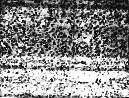
Microscopic examination in all cases disclosed a poorly differentiated small cell tumor with predominantly diffuse areas, but with scattered trabecular and cord-like arrangements. Mitotic activity was high, generally greater than 20 mitotic figures per 10 high-power fields in the most active areas. Necrosis was seen microscopically in 15 cases (75%). Characteristic follicle-like spaces frequently containing pale eosinophilic fluid (Fig. 1 ) were seen in 18 cases (90%). Focal spindle cell areas were present in 6 cases (30%). The tumor margin with residual ovarian stroma was smooth and pushing in all 9 cases in which this interface was visible (45%). A second large cell population was intermixed within the tumor in 14 cases and was seen at the periphery of the tumor in two cases.
lmmunohistochemistry for p53 Protein
Immunoreactivity for p53 protein was detected in 12 of 15 tumors tested (Fig. 2). Staining was focal in 4 tumors and diffuse in 8. Sections from all 9 tumors that were stained within 2 weeks of cutting were immunoreactive (7 diffuse, 2 focal). Sections retrieved from the files 1-3.5 years prior (3 tumors) were positive (2 focal, 1 diffuse), while sections cut 12-14 years previously (3 tumors) were negative.
DISCUSSION
Recently there has been a deluge of reports on the status of the p53 gene in a wide variety of human malignancies. The presence of p53 gene mutations as the most common genetic event in human cancers is established [1, 2]. Of particular importance in the study of p53 gene mutation is the strong correlation of mutation with a prolonged half-life of the abnormal gene product, and thus p53 protein accumulation, allowing for detection of accumulated mutant p53 protein by immunohistochemical methods. Normal low levels of p53 protein are not detectable with this method. In general, there is a strong correlation between p53 protein accumulation and p53 gene mutation, and this has been demŁonstrated for ovarian tumors. However. this correlation is not perfect, and a minority of tumors that accumulate p53 protein do not contain identifiable p53 gene mutations [11, 33].
p53 gene abnormalities have been extensively studied in ovarian carcinomas; p53 mutation or p53 protein accumulaŁtion has been demonstrated in 26-92% of cases [3-16]. Many of these studies have demonstrated a correlation of p53 mutation with increasing stage (FIGO) [3, 5, 13, 15, 16, 33, 34], but not with grade or prognosis. These studies have not included cases of small cell carcinoma of the hypercalcemic type; however, four studies each included one case of undifferentiated carcinoma, not otherwise specified [3, 5, 7, 13].
Small cell carcinoma of the ovary is a highly aggressive tumor of uncertain histogenesis that occurs in young women. It was first described in 1982 [17] and several case reports and small series followed [18-28]; recently, 150 cases were reviewed {29). Studies utilizing immunohistochemistry and electron microscopy have failed to provide convincing eviŁdence of a particular cell of origin [18, 20, 21, 23]. This tumor has been variously argued to be of germ cell or gonadal stromal origin, but at present the evidence is inconcluŁsive [29, 30].
Scully and his associates from the Massachusetts General Hospital have extensive experience with ovarian small cell carcinoma [17, 21, 22, 25, 29, 30]. In the original description, they did not favor a specific origin [17], but in a subsequent immunohistochemical study in which no specific line of differentiation was elucidated [21], they suggested an unusual form of germ cell tumor or a bizarre epithelial tumor. More recently. they found these tumors to be DNA diploid by flow cytometry [22], suggesting a gonadal stromal origin. In their recent review of 150 cases, no new evidence for a particular cell of origin was presented [29].
Although we did not directly analyze the p53 gene, the identification of accumulated p53 protein in 80% of ovarian small cell carcinomas provides data supporting an important role for p53 gene abnormalities in the genesis of this highly aggressive ovarian carcinoma. The absence of detectable p53 protein accumulation only in old (12-14 years) sections of three tumors suggests loss of immunoreactivity with proŁlonged storage in the form of tissue sections and also sugŁgests that virtually all ovarian small cell carcinomas accumuŁlate p53 protein. Because of the strong correlation of staining with tissue storage time, and the dismal prognosis in nearly all patients, no further correlation of clinicopathologic features with prognosis was possible in this material.
The present study also confirms the poor prognosis for patients with ovarian small cell carcinoma. The clinical and pathologic features in our subjects are similar to those deŁscribed in previous reports. Future studies to determine the nature of the p53 mutations in ovarian small cell carcinomas would be of value for further comparison to ordinary ovarian carcinomas.
REFERENCES