|
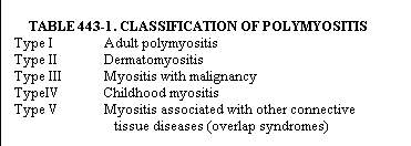
DEFINITION. Polymyositis is an inflammatory disease of skeletal muscle of unknown etiology, characterized by symmetric weakness of the limb girdles, neck, and pharynx. Patients with this illness can be divided into five clinical categories (Table 443-1). In this chapter the term "polymyositis" is used to characterize the whole group of patients, while "adult polymyositis" is used to denote type I patients. INCIDENCE. Polymyositis has an annual incidence of approximately five cases per million population. There is no relationship to birth order, family size, socioeconomic level, or geographic location. Familial cases are unusual. Age distribution is bimodal, with a small peak between ages 10 and 14 and a larger peak around age 50. Patients with myositis associated with malignancy are slightly older, with a mean age just over 60. In adult polymyositis and dermatomyositis, females outnumber males by two to one. In cases associated with overlap syndromes the female dominance is even more pronounced. The sex ratio is equal in myositis associated with malignancy.
PATHOLOGY AND PATHOGENESIS. The characteristic features of polymyositis on muscle biopsy include
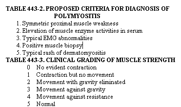
These findings may vary from patient to patient and even on multiple biopsies of a single patient. Approximately 15 per cent of initial muscle biopsies are normal, and the full picture of typical changes can be expected in about 50 per cent. Biopsy should be performed on a muscle that is involved but not totally weakened. Electromyography may aid in localizing an involved area, but the biopsy should not include the exact site of the electromyograph needle puncture, for the needle may cause focal fiber destruction. Biopsy of involved skin shows marked dermal edema, basal vacuolation, and colloid bodies. Similar changes occur in the skin in systemic lupus erythematosus, but in dermatomyositis the basement membrane is normal in thickness and does not stain for immunoglobulin or complement in indirect immunofluorescence studies. The leading theory of the pathogenesis of polymyositis attributes muscle damage to cell-mediated autoirnmunity. Several lines of evidence support this hypothesis. In adult polymyositis approximately one third of the cells invading the muscle are macrophages and two thirds are lymphocytes. The intensity of the inflammatory infiltrate increases from perivascular through the perimysial to the endomysial area. Lymphocytes directly in contact with viable muscle fibers are predominantly cytotoxic/suppressor (T8) in phenotype and often send finger-like projections into the muscle cells. Lymphocytes expressing the helper (T4) phenotype are found in the areas surrounding these invading cells. Studies of the ability of mononuclear cells from polymyositis to kill or damage muscle cells in vitro have yielded conflicting results, but sensitivity of these cells to muscle antigens has been demonstrated both by proliferation and lymphokine production. Soluble mediators released include a factor that inhibits calcium binding by normal sarcoplasmic reticulum. Class I major histocompatibility (MHC) antigens that are crucial to the interaction of T8 lymphocytes with target cells are poorly expressed in normal sarcolemma, but their expression is greatly enhanced in areas adjacent to infiltrating lymphocytes in polymyositis. The increase in Class I MHC antigens is apparently mediated by interferon produced by the infiltrating lymphocytes. Thus, many of the elements needed for cellmediated injury have been identified at the site of pathology in polymyositis. Further evidence for an autoimmune _process in polymyositis comes from the multitude of autoantibodies present in the serum. Antimyosin antibodies occur in 90 per cent and antimyoglobin antibodies in 70 per cent of patients. While not specific for polymyositis, their prevalence is considerably higher than in other muscle diseases. Evidence for antibodies against structures on the surface of muscle cells is less plentiful, but IgG from polymyositis sera can accelerate degradation of acetylcholine receptors of cultured muscle cells. Antinuclear antibodies demonstrated by immunofluorescence on human tissue culture cells (HEp-2) or by precipitation with calf thymus nuclear extract occur in 90 per cent of patients. Specificities for a variety of discrete nuclear components have been identified. Two antibodies, anti-Jo-1 and PL-7, react with histidyl-tRNA synthetase and threonyl-tRNA synthetase, respectively. Anti-Jo-1 antibodies are found in 30 per cent of type I and less than 10 per cent of type 11 patients but in 50 to 100 per cent of patients with adult polymyositis and interstitial lung disease. It is postulated that these unusual antibodies may be the footprints of a preceding viral infection. Certain enteroviruses can mimic tRNA and interact with aminoacyl-tRNA synthetase enzymes. This interaction might render the enzyme immunogenic and account for the presence of the autoantibodies. Anti-PM-Scl antibodies react with a nucleolar antigen and anti-Ku with a novel non-histone DNAbinding protein. Both are found in patients with polymyositis and scleroderma overlap. Anti-Mi-1 and Mi-2 antibodies react with nuclear components and occur in 3 and 20 per cent of dermatomyositis patients, respectively. A small number of polymyositis patients also have antibodies to nRNP, and almost two thirds of the sera react with intermediate filaments of the cytoskeleton. Anti-Jo-1 is associated with HLA-DR3, suggesting a genetic basis for the expression of at least some of these autoantibodies. Vascular damage is also implicated in the pathogenesis of polymyositis, particularly in children, and in dermatomyositis. In a subset of children with dermatomyositis, cutaneous ulceration, and intestinal infarction, the clinical picture suggests necrotizing vasculitis. Examination of tissue, however, reveals primarily noninflammatory endarteropathy and lymphocytic perivasculitis. The terminal membrane attack complex of the complement system is deposited, bound, and activated to completion in muscle microvasculature in most patients with childhood myositis and a smaller portion of adults with dermatomyositis. Correlation with immunoglobulin deposition is poor, suggesting an antibody-independent mechanism of complement activation. In addition, replication of capillary basement membrane and alterations in endothelial cells are frequently seen on electron microscopy. Loss of capillries also occurs. It characteristically starts in the periphery of the fascicles and is more pronounced in children than in adults. These changes are probably not due to muscle atrophy, because the ratio of capillary lumina to muscle cell area does not change in Duchenne dystrophy and increases in denervation atrophy. Infection caused by a virus or an organism such as Toxoplasma gondii has been postulated as a cause of polymyositis. The prevalence of IgM anti-Toxoplasma antibodies is increased in patients with polymyositis, and Toxoplasma organisms have been identified in a few cases of adult polymyositis, but treatment for toxoplasmosis has had little effect on the muscle disease. These cases may represent secondary infection with Toxoplasma. Coxsackievirus has been isolated from a few cases of polymyositis, and the prevalence of antibodies to Coxsackievirus B is increased in children with dermatomyositis. The recently described myositis that occurs in newborn mice after injection with Coxsackievirus BI might provide a system to study the role of both infection and cell-mediated immunity in muscle damage. CLINICAL MANIFESTATIONS. Weakness of the proximal muscles occurs in nearly all patients and is the presenting complaint in 93 per cent. The typical case begins with gradual onset of weakness in the hip girdle and proximal leg muscles. Muscle pain and tenderness are absent or mild. Weakness of the shoulders, proximal arm muscles, and neck flexors follows. Involvement of the pharyngeal muscles may occur with dysphagia, dysphonia, and dysarthria. Early symptoms include inability to rise from a low chair, to climb stairs without the use of a railing, or to raise the arms above the head to comb the hair. In advanced cases, the patient may be unable to lift the limbs against gravity. Muscular wasting is variable and frequently minimal until late in the disease, Contractures are almost exclusively associated with long-standing disease. The ocular muscles are almost never involved, and weakness of distal muscles occurs in less than 20 per cent of cases. Asymmetric weakness, weakness of isolated muscle groups, and acute onset with global weakness are unusual. Deep tendon reflexes are normal or slightly reduced. Muscle symptoms of childhood polymyositis are similar to those of the adult form, but fever, weight loss, rash, contractures, and subcutaneous calcification are more common. Arthralgias occur in about one quarter of patients with adult polymyositis or dermatomyositis. True arthritis is usually mild; begins prior to or coincident with weakness; involves the hands, wrists, and knees; and responds quickly to steroid treatment. Synovial fluid has good viscosity and mononuclear cells. Synovial biopsy reveals fibrin deposition, focal loss of lining cells without proliferation, and mild inflammation. Patients with myositis and overlap syndromes may have joint involvement typical of rheumatoid arthritis or systemic lupus erythematosus. A peculiar arthritis of the hands with erosions, periosteal calcification, and instability of the interphalangeal joints of the thumb has also been described. An erythernatous skin rash occurs on the forehead, neck, shoulders, trunk, and arms of about one third of patients and distinguishes those with dermatomyositis, A lilac or heliotrope rash occurs on the upper eyelids and face in 15 per cent. The rash may be associated with edema. Reddened, elevated, scaly patches are characteristically seen over the extensor surfaces of the small joints of the hands (Gottron's sign), the elbows, the knees, and the medial malleoli. Nailfolds may show periungual telangiectasia, dilated and distorted nailfold capillary loops alternating with avascular areas, or thickening and roughening without redness. In some patients, the finger pads become shiny and atrophic with constant peeling. Patients with myositis and overlap syndromes may display the whole spectrum of dermatologic changes associated with connective tissue diseases. Raynaud's syndrome occurs in one half of the patients with overlap syndrome and one fifth of those with adult polymyositis and dermatomyositis. It is less common in children and in patients with malignancy. Dysphagia in polymyositis is primarily due to weakness of the striated musculature of the posterior pharynx. Dysphagia may also result from cricopharyngeal obstruction secondary to inflammation or fibrosis of the cricopharyngeus muscles. In this case, surgical division of the involved muscles provides prompt relief of the symptoms. Dysfunction of the esophagus and stomach occurs but is usually overshadowed by pharyngeal dysfunction. Hypornobility and poor absorption in the small intestine have been seen in a few patients without symptoms of frank scleroderma. Vasculitis associated with the childhood form of the disease may lead to mesenteric thrombosis. Asymptornatic electrocardiographic abnormalities are common in polymyositis, but cardiac involvement manifested by congestive heart failure or symptomatic heart block occurs in only 10 per cent of patients. Inflammatory cardiomyopathy, fibrosis, and small vessel disease have been found in some of these patients at autopsy. Interstitial pneumonitis occurs in 10 to 15 per cent of the patients. Cough and dyspnea precede muscle weakness in one half of the cases of interstitial pneumonitis. No relationship is apparent between the severity of the lung and muscle involvement. Vasculitis is not characteristic of this lesion, and pleurisy is uncommon. The presence of active inflammation on lung biopsy correlates well with steroid responsiveness. Renal involvement is rare. When renal failure occurs, it is usually attributable to myoglobulinuria. About 13 per cent of patients with adult-onset polymyositis have coexistent malignancy. Females are affected as frequently as males. Several recent studies have found an equal incidence of malignancy in adult polymyositis and dermatomyositis, but the combined data of large series in the past 10 years suggest that patients with dermatomyositis are at greater risk. Myositis precedes or occurs simultaneously with the diagnosis of malignancy in two thirds of patients, and in most instances the two diagnoses are made within the span of a year. Tumors of the breast and lung are the most common. Those of the ovary and stomach occur more frequently than in the general population, whereas tumors of the colon and rectum are less frequent. Polymyositis associated with malignancy has no clinical features that differentiate it from polymyositis alone. Extensive undirected radiographic screening of these patients for cancer has proved unrewarding. Clues to the coexistence of a malignancy are almost always present on history, physical examination, or routine laboratory tests. CLINICAL COURSE AND PROGNOSIS. The five-year survival rate is approximately 80 per cent. The best survival rate occurs in children. Diagnosis at age 45 or above and the presence of cardiac involvement are associated with shorter survival. Death within the first year is often from pneumonia associated with dysphagia and aspiration. The leading causes of death are malignancy, infection, and cardiovascular disease. About half of the surviving patients will attain almost complete recovery of muscle strength. LABORATORY DATA. Serum levels of muscle-derived enzymes, principally creatine kinase (CK), transaminases (SGOT, SGPT), lactate dehydrogenase (LDH), and aldolase, are elevated at some time during the course of the disease in 99 per cent of the patients and will be normal at any one time in about 10 per cent. CK and aldolase are the most sensitive, but it is wise to obtain all five enzymes on initial screening. The MB isoenzyme of CK is occasionally elevated in the absence of cardiac involvement owing to its increase in regenerating muscle. The levels of these enzymes correlate reasonably well with disease activity and can be used as guides to therapy. Approximately one half of patients have elevated serum levels of myoglobin or an abnormal erythrocyte sedimentation rate. Neither test can be relied upon as an index of disease activity. With few exceptions, complement studies are normal. Radionuclide scanning with 19-Tc-polyphosphate may reveal increased uptake in areas of active myositis. The electromyogram (EMG) is abnormal in almost all patients. The most common abnormalities, reduction in amplitude and duration of motor unit potentials, occur in 90 per cent of cases but are not specific for polymyositis. Evidence of membrane irritability, including fibrillation, positive sharp waves, and increased insertional activity, occurs in one half to three quarters of the EMG's. Spontaneous bizarre highfrequency discharges are also characteristic. Electromyographic patterns do not differ among the clinical types of disease. In some patients the characteristic pattern is present only in certain muscle groups. The paravertebral musculature is frequently involved and should be included in diagnostic electromyography. DIAGNOSIS AND DIFFERENTIAL DIAGNOSIS. In order to standardize diagnosis of these patients a set of criteria has been proposed (Table 443-2). The reliability of the diagnosis depends upon the number of criteria met. Patients are classified as having definite disease with four, probable disease with three, and possible disease with two criteria. The presence of proximal muscle weakness is established both by the history and by objective testing. One scheme for grading muscle strength utilizes a five-point scale (Table 443-3). This scheme works well with severe weakness but is relatively insensitive to clinically important changes in the range of grades 4 and 5. A test using 10 timed chair stands offers a reproducible quantitative method to assess lower extremity strength in this critical area.
Polymyositis is but one of a variety of diseases that may be present as muscle weakness with or without muscle pain. Neurologic disease is a primary concern in the evaluation of these patients. The history and physical examination will usually establish the neurologic origin, and EMG will show neuropathic changes. In muscular dystrophy a family history is often present and the symptoms progress over years rather than months. The CK may be elevated and may decrease on treatment with corticosteroids, but clinical improvement does not occur. The early involvement of the ocular and facial muscles helps differentiate myasthenia gravis. Steroid myopathy begins insidiously in the proximal leg and hip muscles and spreads to involve the shoulders and arms. Other signs of glucocorticoid excess are usually present, but there is a poor correlation between the actual dose of corticosteroids and this syndrome. Raising the dose over a previously tolerated level may induce myopathy, and decreasing it to the previous level may relieve the symptoms. The combination of elevated urinary creatine and normal serum enzyme activities has been suggested as a useful differential point in favor of steroid myopathy. Other drugs that may cause myopathy include alcohol, clofibrate, penicillamine, azathioprine, phenytoin, polymyxin, chloroquine, and emetine. Both hyper- and hypothyroidism may cause proximal muscle weakness. In hypothyroidism, serum creatine kinase may reach very high levels, and the electromyogram may mimic that in polymyositis. Hyperparathyroidism may also cause proximal muscle weakness, an abnormal electromyogram, and muscle atrophy without an inflammatory infiltrate. Weakness and myalgia. may occur after exercise in McArdle's syndrome, carnitine palmityl transferase deficiency, myoadenylate deaminase deficiency, or renal tubular acidosis. Acute exertional rhabdomyolysis is characterized by muscular pain, swelling, and weakness and is associated with heme pigment in the urine. It occurs after strenuous exercise in apparently healthy individuals. Infectious causes of chronic myositis include trichinosis and tropical pyomyositis. In the latter, staphylococcal abscesses deep within muscles cause subacute pain and firm swelling, primarily in the gluteal, quadriceps, or trunk muscles. Serum muscle enzymes are normal, and blood cultures are usually negative. Inclusion body myositis is a rare disease that causes slowly progressive weakness of both proximal and distal muscles, usually most pronounced in the lower extremities. It affects primarily middle-aged men, and the response to steroids or immunosuppressive therapy is poor. Diagnosis is made on muscle biopsy, which reveals eosinophilic inclusions in both the nucleus and sarcoplasm of muscle cells. Other causes of muscle weakness and pain include the hypereosinophilic syndrome, diffuse fasciitis with eosinophilia, microemboli from atheromatous plaques or nonbacterial endocarditis, diabetic amyotrophy, and phosphate depletion secondary to the use of nonabsorbable antacids. Polymyalgia rheumatica occurs in the elderly and is characterized by proximal muscle pain and stiffness (see Ch. 442). The only laboratory abnormality is a striking elevation of the erythrocyte sedimentation rate. The absence of muscle weakness and prompt relief after treatment with a small dose of corticosteroid differentiate this disease from polymyositis. Other connective tissue diseases may have myositis as part of the symptom complex. These patients should be labeled as having polymyositis only if they meet the independent criteria for the diagnosis. TREATMENT. Bed rest is necessary during the active stage of the disease. Active physical therapy should be reserved until the inflammation has subsided. In spite of a lack of adequately controlled therapeutic trials, corticosteroids are generally accepted as the drug of choice. Prednisone, 50 to 100 mg, is given daily in divided doses and continued until definite improvement occurs. Serum enzymes typically decrease to half their pretreatment values one month after initiation of therapy and reach normal values in two to three months. Muscle strength usually shows definite improvement in two months. Attempts to decrease steroid dosage rapidly or discontinue treatment prematurely may lead to a recurrence of the disease. A single daily dose or alternate-day steroid therapy may be tried in order to decrease the side effects but should be attempted only when the disease is under good control. Maintenance therapy will be necessary for years in many cases. Failure to respond to steroids occurs in about 20 per cent of patients. Immunosuppressive drugs such as methotrexate and cyclophosphamide, or plasma exchange, may be beneficial in these instances, but controlled studies to substantiate their efficacy are not available. Combined therapy with azathioprine and prednisone has been shown to reduce steroid requirements and improve function in long-term treatment compared with steroid alone. |
ENTRANCE | HOME | 1 | 2 | 3 | 4 | LINKS | FUN STUFF | BULLETIN BOARD | BOOK STORE | DISEASES | SEARCH