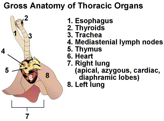

1. Time to start with the basics. You have the heart, thymus, lungs, thyroid, esophagus, larynx, mediastinal nodes, and trachea in front of you in one nice little package. Of course, all need to be examined.
2. Remove the esophagus from the system and check for any blockage by inserting a syringe in one end and flushing with saline or preservative.
3. The thyroids are flanking the trachea on the anterior end of the system and should have a nice tannish color. Check to see if the color is consistent and they are symmetric in size and shape. For those of you who are wondering about the parathyroids, they are not visible grossly at this point. Located around or within the thyroids, these little guys can only be seen histologically.
4. The thymus lies directly on top (or ventrally) to the heart. In younger animals that haven't gotten past that awkward adolescent age, the thymus is going to be rather large. As the animal ages, the thymus shrinks in size. It should be a nice whitish-translucent color without any inconsistencies in color. In athymic mice, obviously there is no thymus. So, don't get all excited if you can't find a thymus on an immunosuppressed mouse.
5. The mediastinal nodes lay dorso-lateral to the thymus and can be enlarged. Clever little lymph nodes that they are, when enlarged they may look like thymic lesions. So be careful!!!!!

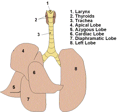
Time to examine the lungs. Using a syringe full of preservative, insert the needle into the trachea and fill the lungs. Not only does this preserve the lungs in a way that is beneficial to histology, but also it will give you a 3 dimensional area to examine for possible lesions. The lungs should have a nice pink bubble gum color. The surface should be smooth and fresh looking. Examine each lobe (see diagram) to see if any lesions are lurking in junctions or hidden from view. Place the whole structure in fixative. The lungs are not as dense as the fixative and will float on the surface. If for some reason, the lungs sink, something is wrong and it should be noted. The only thing that you removed in this whole process is the esophagus and the lungs, heart, thyroids, trachea, thymus, and mediastenial nodes have remained together.
