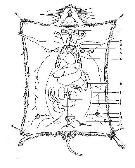
Diagram Key
1. Superficial cervical nodes
2. Deep cervical nodes
3. Mediastenial nodes
4. Axillary node
5. Brachial node
A. Thymus
B. Spleen
6. Pancreatic node
7. Renal nodes
8. Mesenteric node
9. Inguinal node
10. Lumbar nodes
11. Sacral Node
12. Sciatic node
Lymph Nodes
You have reached one of the more frustrating experiences you will ever encounter, trying to find those fun-loving lymph nodes. But alas, those of you who are patient will reap all the rewards of this little exercise. At this point, you are going to be looking for lymph nodes that are located subcutaneously or in the connective tissues between muscles. If there is some type of immune response before the time of the necropsy, it will not be difficult to locate the nodes. On the other hand, if the animal is healthy, wish yourself luck and hope you have good eye sight, because they may be difficult to see. What you are looking for are bead shaped structures about the size of a single grape nut. They can range in color from a yellowish white to tan depending on the strain of the mouse. Obviously, if the mouse is pigmented, the nodes will be darker than that of an albino mouse. Lay the nodes flat on a piece of paper towel and place in fixative. The internal (deep) lymph nodes (see table for locations of selected lymph nodes) lie within the body cavity and can be retrieved at various choice times during the necropsy.
Lymph Node |
Location |
Lymph Node |
Location |
| axillary | Armpit | popliteal | Behind the knee, between the adductor muscle and the semimembranous muscle |
| brachial | Resting on the bicep muscle, underneath the pectorals | pyloric (pancreatic) | Anterior end of the pancreas, near the pyloric end of the stomach |
| deep cervical | Buried behind the salivary gland | renal | Between aorta and kidneys |
| inguinal | Adherent to the skin of the groin | sacral | Posterior to the bifurcation of the abdominal aorta |
| lumbar | Anterior to the bifurcation of the abdominal aorta | sciatic | Below sciatic nerve on back |
| mediastinal | Thymic region | superficial cervical | Anterior to the salivary gland |
| mesenteric | Mensentery of the small intestine and pancreas |
Diagram of Mammary Tissue
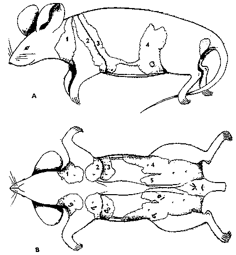 Key
Key
Diagram A
1. Mammary Gland-Left Cervical
2. Mammary Gland-Left Thoracic
3. Mammary Gland-Left Thoracic
4. Mammary Gland-Left Abdominal
Diagram B
1. Mammary Gland-Left Cervical
2. Mammary Galdn-Left Thoracic
3. Mammary Gland-Left Thoracic
4. Mammary Gland-Left Abdominal
5. Mammary Gland-Left Inguinal
6. Mammary Gland-Right Cervical
7. Mammary Gland-Right Thoracic
8. Mammary Gland-Right Thoracic
9. Mammary Gland-right Abdominal
10. Mammary Gland-Right Inguinal
Also, at this time you may wish to examine the mammary tissue of the animal. If the animal is pregnant or lactating the tissue is readily identifiable. It will look as if cottage cheese has been placed between layers of epithelium on the internal surface of the skin. If your animal is not "in the family way" but still looks like mascot for the dairy industry, there might be a pituitary lesion to blame. A nonpregnant, nonlactating, or male mouse should not have a great deal of visible mammary tissue. In those genetic manipulation studies aimed at studying mammary tumors, the tumors will be quite obvious clincially.
