Lymphocytes Polarize Before They Crawl

vimentin and plectin redistribute to the uropod after SDF-1a stimulation. Click for Brown et al PubMed
Shaw Lab

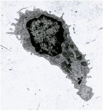
Complex and elegant shapes are  assumed by lymphocytes turning (left) or migrating with trailing uropods (below). Click on any image for more information.

PubMed - Microtubule retraction into the uropod and its role in T cell polarization and motility. Ratner et al

Lymphocytes circulating in the blood are normally spherical and covered with microvilli. Activated to migrate by chemokine, a lymphocyte polarizes its cytoskeleton and organelles as it retracts its microvilli. An array of microtubules and 10nm vimentin filaments create cell rigidity that stabilizes shape or inhibits cell movement. Ratner proposes, that removal of microtubules to the uropod releases cells from inhibition. Betsy Repasky finds that Protein kinase C and Spectrin move from the cell periphery to the uropod; and, the Shaw Lab finds that vimentin, plectin and fodrin also go to the uropod quickly in response to chemokine signals directing cell polarity and movement. Cytoskeletal polarization seems to be aligned so that acquisition of signal and start of movement are correctly at opposite ends of the cell. Nieto et al saw initial expression of chemokine receptors is at the leading edge. Adhesion receptors are first expressed there also, but move to the uropod upon migration.

Click for Lymphocyte Cytoskeleton - HEV migration paper by Anderson et al
See cells fixed while migrating Lymphocytes Polarize as they cross HEV into lymph nodes

These Images illustrate the polarized shape of lymphocytes frozen in the act of locomotion. The three black and white electron micrographs are unpublished data from Adv. Exp. Med & Biol 114:73-83,1979 [PDF]. They show that microtubules are deployed into the uropod as migration begins. In Ratner's confocal image actin concentrated in the anterior lamellipodium is green and bundles of tubulin in the posterior uropod are red. Vimentin and plectin also redistribute to the uropod after SDF-1a stimulation, thus cytoskeletal polarization seems to be aligned so that acquisition of signal and initiation of a motor response toward the signal is coordinated by cell membrane receptor cytoskeletal interactions.


Home Page | Recirculation | Emigration at HEV | FRC Conduit | Chemotaxis & Links

Dendritic Cells in TDL | Dendritic cells in Tissue | Immunology Lecture Notes