

OK, I'm not a zoologist or biologist, but quite often I'm asked more
general snail questions. Not having any knowledge of my own of this,
I've trawled the books and come up with this little page of information, which to
the best of my knowledge is as correct as can be ......
The shell
Shells are composed of several different layers. The thin outer layer (periostracum) consists of an organic substance known as conchiolin. Below the conchiolin there is a much thicker inorganic layer composed of three layers of crystalline calcium carbonate which was formed on an organic matrix. The innermost layer nacre (or mother-of-pearl), is best seen in marine molluscs, only traces of it are to be seen in 'non-marine' molluscs.
The growth of the shell is not completely regular and even. Shell formations are influenced by a variety of factors, including growth hormones, food and temperature. The colouring of shells is produced by organic pigments acquired by the animal from its food. The basic colour and design are determined genetically, although variations can develop due to the above factors.
The diagram below shows the parts of the shell:
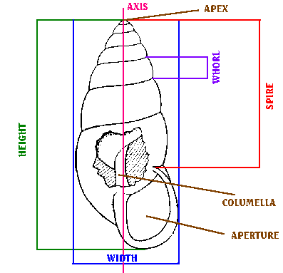
During the life of the snail the whorls of the shell grow steadily wider and wider. When the snails becomes adult, the pattern of growth changes. The shell does not get larger, but the area around the mouth may be strengthened with a rib or lip.
The Snail
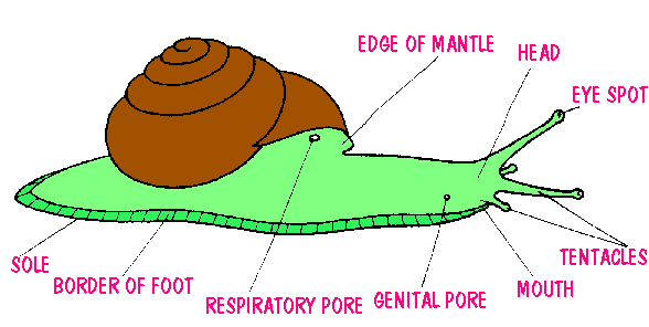
The bodies of slugs and snails are soft and wet, being coated in a layer of mucous (or slime!). The body consists of a bilaterally symmetrical foot and head, and a coiled, asymmetrical hump that fits into the spire of the shell.
Movement
The foot, extruded from the shell, is used for locomotion, which normally takes the form of regular, continuous gliding. It is composed of string muscles, and is connected antieroly with the head, which has a mouth and sensory organs. The ventral surface of the foot is known as the sole, which is surrounded by the foot fringe (or border). The sole slides over a thin layer of mucous secreted by a gland situated in the anterior part of the foot. The picture below shows the foot, and this avi will show the snail in motion, and you can see how these muscle 'waves' work. The thick skin on the back and sides of the body also contains large numbers of mucous glands. Mucous released in to the network of furrows between the tubercles (small bumps on the skin) spreads over the whole of the animal's body, so that the evaporation of water from the animal's skin is reduced.
The digestive system begins on the head with the mouth, continues with the oesophagus, crop, stomach and intestine, which is folded in loops, and terminates in the anus, at the front of the mantel cavity.
Gastropods have an open blood system with blood spaces instead of veins (ie the blood circulates in the general body cavities). The heart, enclosed in a think pericardium, consists of a single thick-walled ventricle and a single thin-walled auricle (atrium). The blood takes up oxygen from the capillary network of the lung and then transports it, via the pulmonary vein, to the atricle from there to the ventricle. The ventricle expels the oxygenated blood into the arteries and after it has also passed from the arterioles it bathes the organs tissues directly, which obtain oxygen and food from it and discharge their waste products: the blood returns to the lung by a series of blood spaces rather than veins.

The most powerful muscles are those of the foot, particularly in the region of the sole, where they are used for locomotion. Another powerful muscle, attaching the snail's body to the internal spiral of the shell, is the columellar muscle, which is inserted on the middle and sends branches to the head and tentacle sheaths which retract the tentacles. By contracting the columellar muscle the snail withdraws into the shell.
The Head
The head is not distinctly separate from the rest of the snail's body. Land snails have two pairs of retractile tentacles, upper pair (ie. the posterior pair) and the shorter, lower pair (ie the anterior pair). The eyes are located on the longer, upper pair.
These avi's show the snail's eyes retracting:
1. Snail retracting eyes (1012kb)- this is a side view of a snail pulling it's eyes in.
2. Snail retracting eyes (706kb)- this is a front view of a snail pulling it's eyes in.
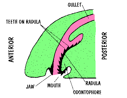
The mouth is partially covered by a pair of adoral lobes, or lips, but the jaw on the upper lip can be seen when the animal is feeding. , this scrapes and cuts the food. On the underside of the oesophagus is a protruding structure known as the radula (or tongue), the convex surface of which is covered with a fine chitin membrane and a quantity of minute teeth arranged in transverse and longitudinal rows. The radula rubs the food against the jaw like a rasp or greater. Although the anterior end of the radula is constantly being worn down, and old teeth broken off and discarded, it's posterior end never stops growing, providing a continual supply of new teeth. The diagram below (vertical section through the head) shows the snail's mouth:
These avi's will show a snail eating:
1. Side view of snail munching lettuce (1867kb)- this is a side view of a snail eating some lettuce, here you can see the mouth movement.
2. Snail munching lettuce (1852kb)- front view of snail eating lettuce.
3. Snail eating lettuce again! (1670kb)- though on this one you can really see the way the snail uses it's mouth to grate away at the food.
Reproduction
Pulmonate gastropods are hermaphrodite and their reproduction is complex. The gonad is an ovotestis, producing both sperm and eggs. The duct of the gonad is in most cases divided, with one channel for the transport of sperm, and the other for the eggs. Furthermore, pulmonates have accessory albumin and mucous glands and, in the oviduct of some families, a special evagination or dart sac. A thin, sharp-pointed chalky structure known as a love dart is formed in this sac and during mating the partners thrust it into each other as stimulation just before exchanging sperm. Although they can function as both male and female, the sperms are formed earlier than the eggs.
This picture shows a snail's dart about to appear:

Here's an avi of a snail getting ready to mate (1343kb)- this is a side view of a snail showing the genital opening (the white dot on the side of it's head, by the eye). This is the movement it'd make on meeting a suitable mate.

Mating is preceded by courtship, during which the partners circle one another, touching one another, and leaving a large platform of accumulated slime. Eventually the love darts are discharged, these may protrude for a while through the atria, or they may actually lodge in the body of the partner. During mating itself, which may last several hours, the snails align themselves so that their common reproductive organs are contiguous; the penis of each is everted and inserted into the vagina of the partner. Sperm is transferred in a long, thin package, the spermatophore, which is formed in the flagellum beyond the epiphallus. The intact spermatophore is passed into the blind diverticulum of the spermatheca duct of the partner receiving it, where it is broken down. The liberated sperm are stored in the spermatheca until required. They then swim up the oviduct, and fertilise the eggs in the region of the albumen gland. Sperm may be stored for more than a year, but egg laying usually takes place about a fortnight after mating.
Updated 14 April 2000
Text and photos © copyright Annette K Goodman, 1998/99/00/01/02/03/04. Any
part of this information may be printed off and used by individuals for non-profit purposes only, as long as the appropriate
acknowledgement of authorship is noted.