What is a
Hypothalamic
Hamartoma?
An Expert Speaks
Out About HH
by Dr Kore Liow, of Kansas University
The Endocrine System
Learn more about the importance of
the endocrine system, hormones and the hypothalamus
MRI Scans of
"The Real Deal"
Actual MRI film of HH
Medical
Articles
Check out our list of
references on HH & Gelastic Seizures, Precocious Puberty and articles on HH and
surgery
See a full description of
Seizure
Types
Back to
HHUGS Home Page
|
|
The following information has
been taken from the interesting pages
at: http://www.psychiatry.wisc.edu/courses/psych619/text/Ch03.html
(for more information about the hypothalamus
click here)
The tiny hypothalamus serves as the Health Maintenance Organization of
the body, regulating its homeostasis, or stable state of equilibrium. The
hypothalamus also generates behaviors involved in eating, drinking,
general arousal, rage, aggression, embarrassment, escape from danger,
pleasure and copulation. It does an amazing number of housekeeping chores
for such a small piece of tissue. Its lateral and anterior parts seem to
support activation of the parasympathetic nervous system: drop in blood
pressure; slowing of pulse; and regulation of digestion, defecation,
assimilation, and reproduction in such a way as to contribute on the whole
to rest and recovery. The medial and posterior hypothalamus regulate
activation: acceleration of pulse and breathing rates, high blood
pressure, arousal, fear and anger.Stimulation of specific groups of cells in these areas can elicit pure
behaviors. For example, rats placed in an experimental situation where
they can press a lever to stimulate a pleasure center will do so to the
exclusion of eating and drinking. Stimulation of another area can produce
rage.
1. Hypothalamus = Homeostasis
The main function of the hypothalamus is homeostasis,
or maintaining the body's status quo. Factors such as blood pressure, body temperature,
fluid and electrolyte balance, and body weight are held to a precise value called the
set-point. Although this set-point can migrate over time, from day to day it is remarkably
fixed.
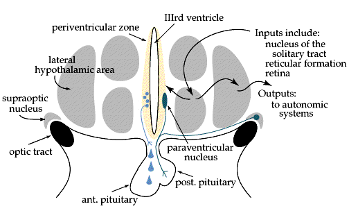
To achieve this task, the hypothalamus must receive inputs about the state of the body,
and must be able to initiate compensatory changes if anything drifts out of whack. The
inputs include:
- nucleus of the solitary tract - this nucleus collects all of the visceral sensory
information from the vagus and relays it to the hypothalamus and other targets.
Information includes blood pressure and gut distension.
- reticular formation - this catchall nucleus in the brainstem receives a variety
of inputs from the spinal cord. Among them is information about skin temperature, which is
relayed to the hypothalamus.
- retina - some fibers from the optic nerve go directly to a small nucleus within
the hypothalamus called the suprachiasmatic nucleus. This nucleus regulates
circadian rhythms, and couples the rhythms to the light/dark cycles.
- circumventricular organs - these nuclei are located along the ventricles, and are
unique in the brain in that they lack a blood-brain barrier. This allows them to monitor
substances in the blood that would normally be shielded from neural tissue. Examples are
the OVLT, which is sensitive to changes in osmolarity, and the area postrema,
which is sensitive to toxins in the blood and can induce vomiting. Both of these project
to the hypothalamus.
- limbic and olfactory systems - structures such as the amygdala, the hippocampus,
and the olfactory cortex project to the hypothalamus, and probably help to regulate
behaviors such as eating and reproduction.
The hypothalamus also has some intrinsic receptors, including thermoreceptors
and osmoreceptors to monitor temperature and ionic balance, respectively.
Once the hypothalamus is aware of a problem, how does it fix it? Essentially, there are
two main outputs:
- neural signals to the autonomic system - the (lateral) hypothalamus projects to
the (lateral) medulla, where the cells that drive the autonomic systems are located. These
include the parasympathetic vagal nuclei and a group of cells that descend to the
sympathetic system in the spinal cord. With access to these systems, the hypothalamus can
control heart rate, vasoconstriction, digestion, sweating, etc.
- endocrine signals to/through the pituitary - recall that an endocrine signal is a
chemical signal sent via the bloodstream. Large hypothalamic cells around the third
ventricle send their axons directly to the posterior pituitary, where the axon
terminals release oxytocin and vasopressin into the bloodstream. Smaller
cells in the same area send their axons only as far as the base of the pituitary, where
they empty releasing factors into the capillary system of the anterior pituitary.
These releasing factors induce the anterior pituitary to secrete any one of at least six
hormones, including ACTH and thyroid-stimulating hormone (TSH).
Ultimately the hypothalamus can control every endocrine gland in the body, and alter
blood pressure (through vasopressin and vasoconstriction), body temperature, metabolism
(through TSH), and adrenaline levels (through
ACTH).
In the news lately:
The hypothalamus controls body weight and appetite, but it is not entirely clear how.
Sensory inputs, including taste, smell, and gut distension, all tell the hypothalamus if
we are hungry, full, or smelling a steak. Yet it is mysterious how we are able to vary our
eating habits day to day and yet maintain about the same weight (sometimes despite all
efforts to the contrary!) . The "set-point" theory is an old one in diet
science, but until recently the mechanics of maintaining that set point were unknown. It
appears that there is an endocrine component to the appetite system. Recent studies in
mice have shown that the fat cells of normal overfed mice will release a protein called leptin
(or OB, after the gene name), which reduces appetite and perks up metabolism.
Leptin is presumably acting on the hypothalamus. Underfed mice, on the other hand, produce
little or no leptin, and they experience an increase in appetite and a decrease in
metabolism. In both of these mice, the result is a return to normal weight. But what would
happen if a mouse (or human) had a defective OB gene? Weight gain would never trigger fat
cells to release leptin, the hypothalamus would never slow the appetite or increase
metabolism, and the mouse would slowly but surely become obese (how the gene got its
name). Sure enough, shortly after these experiments hit the news, the human OB gene was
discovered and a few obese patients were found to have the mutation. Many more obese
patients had normal OB genes, however, indicating that there is much more to the story yet
to be discovered.
2. The anatomy of the hypothalamus
The hypothalamus, as you would expect from the name, is located below the thalamus on
either side of the third ventricle. These sections have been cut coronally, and show only
one side of the hypothalamus.

In this anterior section through hypothalamus, you can see the large neurons of the
paraven-tricular nucleus, which send axons to the posterior pituitary. The cells in the
periventricular zone send axons to the median eminence, from which releasing factors are
carried to the anterior pituitary. The nucleus basalis is a cholinergic nucleus involved
in sleep and wakefulness.
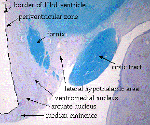
This section is posterior to the first. The hypothalamic nuclei are hard to
distinguish, but the arrows point out approximate locations. The pituitary stalk would
normally be continuous with the median eminence, but it is a fragile structure usually
lost in dissection. Note the fornix descending through the hypothalamus. The fornix
originates in the hippo-campus and ends in the mammillary bodies.

In this posterior section you can see the fornix joining the mammillary body. This is
also a nice section to demonstrate the way that the internal capsule fibers flow into the
cerebral peduncle.
3. The autonomic nervous system
The autonomic nervous system is an entire little brain unto itself; its name comes from
"autonomous", and it runs bodily functions without our awareness or control. It
is divided into two systems which, where they act together, often oppose each other: the sympathetic
and parasympathetic systems. The sympathetic system evokes responses characteristic
of the "fight-or-flight" response: pupils dilate, muscle vasculature dilates,
the heart rate increases, and the digestive system is put on hold. The parasympathetic
system has many specific functions, including slowing the heart, constricting the pupils,
stimulating the gut and salivary glands, and other responses that are not a priority when
being "chased by a tiger". The state of the body at any given time represents a
balance between these two systems.
The best way to learn the functions and structures of each system is by comparison. The
following table lists some attributes of each:
| The Parasympathetic System |
The Sympathetic System |
Origins: |
|
| Parasympathetic cells are located in different nuclei
throughout the brainstem, as well as a few in the sacral spinal cord. Their
axons travel to the target organ, synapse in ganglia in or near the organ wall, and
finally innervate the organ as "post-ganglionics". Examples of these ganglia
include the ciliary, otic, and pterygopalatine ganglia in the head, and diffuse networks
of cells in the walls of the heart, gut, and bladder. Nuclei of origin:
Edinger- Westphal nucleus - Axons from this nucleus travel with cranial nerve
III and have 2 functions:
- pupil constriction
- lens accommodation
Salivatory nuclei - These nuclei in the medulla send axons to the salivary
glands via the VIIth and IXth nerves.
Dorsal nucleus of the vagus - This nucleus gives rise to the secretomotor fibers
of the vagus nerve (X). Its functions include:
- stimulate gastric secretion
- stimulate gut motility
- stimulate respiratory secretions
Nucleus ambiguus (and surrounding cells) - Axons from these cells project via
the vagus to the heart, lungs, and pharynx. Functions include:
- decrease heart rate
- bronchial constriction |
The cells of the intermediolateral column in the thoracic
spinal cord are the source of all the sympathetics. They also travel to ganglia before
reaching the target organ, but the sympathetic ganglia are often far from the target. Some
notable ganglia: Superior cervical ganglion - supplies sympathetics to the head,
including those that:
- dilate the pupils
- stimulate sweat glands
- lift the eyelids
Celiac and mesenteric ganglia - These ganglia distribute sympathetics to the gut.
Functions include:
- vasoconstriction
- inhibition of secretions
Chain ganglia running along the spinal cord distribute sympathetics to the thorax
and periphery to:
- increase heart rate
- dilate bronchi
- selectively vasoconstrict
- vasodilate in active muscles |
The autonomic system also receives afferents that carry information about the
internal organs. They return to separate locations: |
Parasympathetic afferents
Nearly all of the afferents return via the vagus to a single nucleus, the nucleus
of the solitary tract. Like all sensory afferents, the actual cell bodies of the
neurons sit just outside the CNS in a ganglion (the nodose ganglion). The central
processes of the neurons enter the medulla in the solitary tract and travel a bit
before synapsing in the surrounding nucleus of the solitary tract. The solitary tract is
somewhat analogous to Lissauer's tract in the spinal cord.
The nucleus receives information about blood pressure, carbon dioxide levels, gut
distention, etc. |
Sympathetic afferents
Afferents reenter the dorsal horn of the spinal cord along side of the sensory
afferents from the skin. The sympathetic afferents mainly carry information about visceral
pain. Since this information converges with pain from the body surface, the pain is often
perceived as originating at the body surface instead of deep in the viscera. This
phenomenon is called referred pain, and follows predictable patterns. For example,
afferents from the heart enter the spinal cord at the same level as those from the
shoulder region. This is why pain in the heart (a heart attack) is often referred to the
shoulder. |
4. The baroreceptor reflex
A reflex is a pathway with an afferent signal (sensory) that evokes an efferent
response (motor). The most common example is the stretch reflex, or knee-jerk reflex. A
quick stretch of the tendon causes a brief contraction of the muscle. The autonomic system
has several similar reflexes. One of these is the baroreceptor reflex, which maintains a
constant blood pressure despite standing up or lying down.

The afferent signal comes from baroreceptors in the carotid sinus, a swelling of the
carotid artery in the neck. If blood pressure suddenly jumps up, the baroreceptors respond
and send the signal back to the nucleus of the solitary tract (NTS). Neurons in the NTS
project to an adjacent vagal nucleus, the nucleus ambiguus, and excite the neurons that
project to the heart. These acetylcholinergic neurons slow the heart, bringing down the
blood pressure a little.
However, there is more to the story. In the knee-jerk reflex, for the quadriceps muscle
to contract briefly, the hamstring muscle must also relax briefly. As a flexor-extensor
pair, they must always receive opposite signals. The sympathetic and parasympathetic
systems are like a flexor-extensor pair, so when activating the parasympathetic you must
inhibit the sympathetic. Just like in the spinal cord, this is accomplished by an
inhibitory interneuron.

When the high blood pressure signal arrives at the NTS, an inhibitory interneuron
projects to the group of cells that control the sympathetic neurons in thoracic cord.
These cells are called the descending sympathetics. An important feature of the
descending sympathetics is that they are constantly firing at a steady level. This enables
them to be turned down - if a neuron was already silent, an inhibitory signal would make
no difference. Therefore, in response to the surge in blood pressure, the descending
sympathetics are inhibited, and the sympathetics in the spinal cord fire at a much lower
rate. As a result, the heart and the blood vessels are allowed to relax, the heart slows,
vasodilation occurs, and blood pressure drops. The inhibition of the sympathetic system is
actually a more powerful way to lower blood pressure than activating the parasympathetic
system.
What is a
Hypothalamic Hamartoma?
An Expert Speaks Out
About HH
The
Endocrine System
MRI Scans of
"The Real Deal"
Medical
Articles
Full
Description of Seizure Types
Back to HHUGS Home
Page |
