IVP's
An IVP (short for Intravenous Pyelogram) is a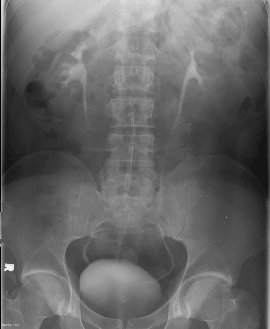 functional study
of the Urinary system. In other words, an IVP demonstrates how
well your Kidneys, Ureters, and Bladder are working in addition
to finding any physical defects or blockages. In terms of what
to expect when you go to have your IVP, the study is fairly simple.
functional study
of the Urinary system. In other words, an IVP demonstrates how
well your Kidneys, Ureters, and Bladder are working in addition
to finding any physical defects or blockages. In terms of what
to expect when you go to have your IVP, the study is fairly simple.
The first thing to take care of is getting yourself prepared for
the examination. Since the urinary system is directly behind the
intestines that make up the largest part of the digestive tract,
it is important that your intestines be reasonably empty. To this
end, some facilities will tell you to eat "low residue"
foods the day before. If the facility at which you are going to
have your study is one of these, your doctor should give you a
sheet with suggestions of foods on it, or you can see the Barium
Enema page on this web site. Most of the organizations I have
worked for however do not require a special diet, instead they
will tell you to drink one bottle of Citrate of Magnesia
(obtainable at most pharmacies and many grocery stores) at 8:00
the night before your exam, and then not to eat or drink anything
except 2 large glasses of water about three hours before the exam
until after you are through.
When you report to the x-ray department for your exam, the technologist
will ask you about your problem (including where you hurt and
if you are in pain now), your medical history (i.e.-have you ever
had a kidney stone, do you have any known kidney problems, etc.),
and whether you are allergic to anything. If you are an outpatient,
they will also have you change into a hospital gown to insure
that there is nothing that might show up in the x-rays. You will
then be taken to the x-ray room where one to three "scout"
films are taken. Just as army scouts precede the army into battle
to find out things the generals will need to know, so too do scout
films precede most contrast studies such as IVP's so that the
technologist and radiologist who will be doing your exam will
not be taken by surprise by unusual problems and your exam delayed.
After the scout films have been taken, the radiologist, a nurse,
or a specially trained technologist will start an IV on you if
you do not already have one. This is to enable the technologist
or radiologist to administer a contrast media that, when concentrated
in the kidneys, will show up on x-rays. You may also be fitted
with a compression band just below the kidneys which, when inflated,
will delay the emptying of the kidneys, thus allowing the technologist
more time to take a special set of x-rays of the kidneys called
tomographs. Tomographs use movement of the x-ray tube and
the film holder (called a bucky) to blur out objects in the body
above, and below, the part we are most interested in. IVP's are
not the only exams this technique is used in, but they are the
most common exam to use it today.
Following three to five tomographs of the kidneys, regular x-rays
are taken at various intervals to watch the contrast containing
urine as it moves down the ureters from the kidneys to the bladder.
Once a certain point is reached, usually 7 to 10 minutes after
the contrast has been administered, two obliques, will
be taken. These x-rays are taken after rolling you, the patient,
about 30 to 40 degrees to your right side, and the same to your
left. These are taken because the ureters enter the bladder low
and towards the rear, so to see the junction of the ureters and
the bladder it is necessary to take an angled shot. The reason
you are the object angled, and not the x-ray tube, is that x-ray
tables are designed so that x-rays coming in to the table at an
angle from the side are blocked. This is to reduce a problem called
scatter that occurs when x-rays are deflected inside the
body in odd directions. If enough scatter can reach the x-ray
film, one ends up with a gray, unreadable mess, so steps are taken
to limit scatter when ever possible. One way to think of this
is to compare it to polarized sunglasses. While the polarization
does not reduce the amount of direct sunlight that gets through
by very much, it greatly diminishes the glare caused by sunlight
bouncing off the pavement and on coming cars.
Once enough contrast, or dye reaches the bladder from both
kidneys, you will be asked to go to the bathroom and empty your
bladder through a special funnel designed to trap kidney stones
for later analysis in the lab. Finally one last x-ray is taken
to see if there is an unusual amount of urine left in the bladder
after you relieve yourself. Some facilities will do this "post
void" film with you standing up in order to make sure that
the kidneys or bladder do not sag or drop, which would indicate
a possible problem with the tissue responsible for holding your
kidneys and/or bladder in the correct position.
Of all the questions I have been asked about this exam, two are
the most common. The first is how long will this exam take. Unfortunately,
this question cannot be answered with any surety, as it depends
entirely upon what is wrong with you. The average healthy patient
with no problems with his urinary system can probably complete
this exam in 15 to 20 minutes. However if something, such as a
kidney stone, is causing a blockage that delays one of the kidneys
from emptying into the bladder, the time required to complete
the IVP can be greatly extended. I once saw a patient who took
so long for the one kidney to empty that the exam was stopped
before we had finished. Yet after treatment in lithotripsy the
next day, this patient was able to be released the same day. So
don't panic if your IVP seems to be taking an extremely long time.
Wait until the doctor explains your problem to you. It may be
that, while bad, it is still nothing to get too concerned about.
The second question I often am asked is how safe is the "x-ray
medicine" or contrast we use. While it is true that the ionic
compounds commonly used 20 years ago did have some problems, the
newer non-ionic dyes commonly used today are extremely safe. In
a study of 2,098 patients who received Optiray, a contrast
media produced by Mallinckrodt Medical, Inc., less than 1% experienced
any reactions that could be considered serious. In addition, all
registered and/or licensed technologists are trained to recognize
the signs and symptoms of any such reaction immediately, and drugs
designed to counteract these reactions are kept in each room where
IVP's may be performed. The most you are likely to experience
from the contrast is a slight flushing sensation, and maybe a
coppery/metallic taste in your mouth. And even these are by no
means sure. Many patients go through this exam without ever experiencing
any sensations at all.
[Home] [X-rays]
[Cat Scan] [MRI] [Mammography] [Links] [Bio]