
 Artigo Original
PATIENTS
Thirtyfour patients underwent pulmonary resections using folded pericardial patchs to reinforce "hand made" parenchymal sutures. The 15 male and and 19 female patients ranged in age from 10 to 75 years
(Mean 48.5). Indications for surgery included various diseases Table 1 and different types of resections were
performed Table 2.
TABLE 1 -
INDICATIONS FOR SURGERY
CAVITARY INFECTIOUS DISEASE n=2
Micetoma(2)
NODULAR DISEASE n=12
Adenocarcinoma(4)
Hamartoma(2)
Extrinsec alveolitis(1)
Tuberculoma(5)
OBSTRUCTIVE DISEASE n=8
Type I Bulla(2)
Type III Bulla+Emphysema(2)
Emphysema(4)
| (N) | Length of suture(cm)* | |
|---|---|---|
| Byopsies | ||
| Wedge | ||
| Atypical Segmentectomy | ||
| Wedge+Biopsy | ||
| Cavernoplasty | ||
| Bullectomy | ||
| Reduction Pneumoplasty |
TECHNIQUE
Pericardial doublet folded patchs®* are tailored from rectangles of low thickness bovine pericardium to fit both grasping claws of an autoesthatic clamp (Rochester type)
(Figure 1).
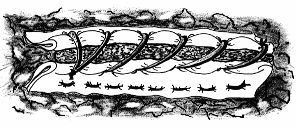
The folded pericardial patchs are than used to sandwich clamping of the lung parenchyma (Figure 2a). Following pulmonary resection an horizontal suture is made using absorbable suture and involving successively pericardium, lung and pericardium (Figure 2b).
(a) (b)
(b)
Figure 2 - (a) Sandwhich clamping of lung and resection;
(b) Horizontal suture underneath the grasping claws of the clamp.
The clamp is then unblocked and gently removed with a retrograde movement(Figure 3).

The procedure is completed with a continuous suture using the pericardial patchs to buttress the sandwiched cutted lung edge (Figure 4).
RESULTS
Immediatly after completion of the suture the "water test" did not show air leaks in 33 of the 34 patients through the suture line
(Mean Length=11.5cm; Range=6.5-24 cm).
In these 33 patients there was no postoperative air leakage and the mean drainage time was 1.3 days (Median=1; Range=1-3) and the mean postoperative
time was 3.9 days (Median=4; Range=2-5)Table 3.
TABLE 3 - RESULTS
| NO AIR LEAK
(N=33) | AIR LEAK
(N=1) | TOTAL
(N=34) |
|
|---|---|---|---|
| Length of Suture (cm) | |||
| Drainage Time (days) | |||
| Postoperative Time (days) |
COMMENTS
Postoperative air leakage is considered a common sequela of pulmonary resections.
When suturing the lung with fine needle sutures a point of diminishing return is reached after which the surgeon accepts a certain amount of leakage. This is better than producing new tears with repeated attempts at sewing over(3).
Stapling is a fine swift way of suturing the lung, though it is not clear if stapling produces a safer air stanching suture, inasmuch as the staples may not be allaways uniformily crimped, situation in which it is necessary the use of complementary safety manual stitches.
In routine pulmonary surgery air leakage of short duration lasting less than 4 days, is tolerated as an inevitable event. Prolonged air leakage lasting more than 5 days do occur after routine resections, however it is mostly common following surgery for emphysematous bullous disease(4).
The recent surgical retake of reduction pneumoplasty for emphysema was made possible safely and with low morbidity by the use of pericardial strips fastened to the stapler and than suited to buttress the suture line. This approach greatly reduced the incidence of air leakage in the fragile emphysematous lung tissue.
The corollary of the effectiveness of stapling with pericardium is the expectation of attainment of perfect results, i.e. "dimininished air leaks" if this technique is used routinely in pulmonary resections, as suggested by Urschell(2).
The folded pericardial doublet patchs that we report here produced extemelly safe sutures. Results shows a dramatic decrease in the occurrence of air leakage after routine pulmonary resections. Air leaks simply did not occurred in 90% of the cases. The overall mean drainage time was 1.7 days and the mean postoperative time was 4.3 days.
These times are significantly lower than those obtained after pulmonary resections using simple surgical sutures and/or stapling (Mean Drainage Time=5.4 days and Mean Postoperative Time=11 days)(5).
The effectiveness of the protection conferred by the folded pericardial patchs was experimentally confirmed performing sutures of "ex-vivo" lungs of dogs. The delicate and fragile dog's lung parenchyma don't accept neither manual nor mechanical sutures. Nevertheless reinforcing the suture with pericardial patchs results in perfectly air stanching sutures.
From a technological and practical point of view stapling with pericardium is the ideal technique due to the clearness and quickness with which the suture is done. However manually reinforcing the suture with pericardium does not depreciates the surgeon's activity for the time expent to perform the suture (10 to 20 minutes for a 15cm suture); time thus expended that represents significant savings in costs to attain an equal clinical benefit, i.e. decreased air leakage, decreased drainage time, decreased postoperative time and a more rapid convalescence.
Our results shows that "hand suturing" the lung with pericardium is a safe alternative technical way of preventing air leakage standing
for prompt and smooth convalescence from pulmonary resections. Incomplete convalescence due to air leakage of any intensity
is especially alarming to the patient and the family, distressing to the surgeon and expensive to the health care system.
So that it is needless further convincement or persuasion to accept that air leakage is a matter of major concern to patients,
doctors and those who pay the bill.
CONCLUSIONS
The use of the folded pericardial patchs technique to manually reinforce the lung suture line, produces safe air stanching parenchymal
sutures, resulting in a dramatic decrease in postoperative air leakage,
thus favoring prompt and smooth convalescence following routine pulmonary resections.
REFERENCES
NOTE:
Functional Improvement After Volume Reduction: Sternotomy Versus
Videoendoscopic Approach
Access the articles:
Prevention of Air Leaks After Pulmonary Wedge Resection
by/Cecil C. Vaughn, Ernst Wolner, Marcel Dahan et al.
Annals of Thoracic Surgery
by/Wilfried Wisser, Edda Tschernko,
Ömer Senbaklavaci et al.
Annals of Thoracic Surgery
(1)Cooper,JD et al.: Bilateral pneumectomy (volume reduction) for Chronic
Obstructive Pulmonary Disease
J. Thora. Cardiovasc. Surg., 1995, 109:106-19.
(2)Urschel,HC.: Bilateral Pneumectomy (volume reduction)[Discussion].
J. Thorac. Cardiovasc. Surg., 1995, 109:106-19.
(3)Vogt-Moykopf,I and Meyer,G.: Management of prolonged parenchymal air leaks and residual pleural spaces [Discussion]. In International Trends in General Thoracic Surgery, WB Sauders Company, 1987,2:392-3.
(4)Connolly, JE and Wilson, A.: The current
status of surgery for bullous emphysema.
J. Thorac. Cardiovasc Surg. 1985;97:351-61
(5)Thévenet, F et al.: Pneumothorax spontané et
récidivant. Traitement chirurgical - a propos de 278
observations.
Ann. Chir. thorac. cardio-vas., 1992;46:165-169

