Bicornuate Uterus
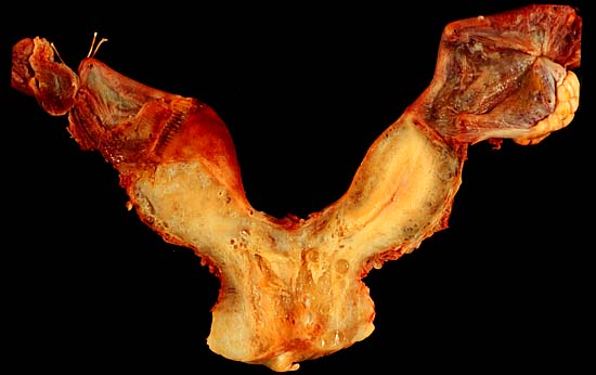
This uterus was removed in the course of excising an ovarian mucinous cystadenoma. The photo above shows the uterus and adnexa (less the initially excised left ovarian tumor) from the posterior aspect. The pale yellow right ovary is clearly seen on the upper right of the image. Both oviducts, visible at the top right and left, adjoin their respective separate cornua of this malformed uterus.
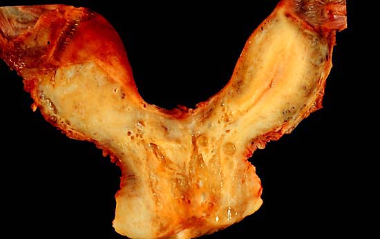
The photo below is a closer posterior view of the uterus. The endocervical canal and common endometrial cavity are visible at the midline. The endometrial cavity of the right cornu can be seen, but the slice misses the full extent of the left cornu's endometrial cavity. Exam with a probe disclosed that both cornua had full-length endometrial cavities.
Although this is a rather dramatic uterine deformity, this patient had carried normal to pregnancies to term. Bicornuate uteri, while rare in humans, are normal in many mammalian species, including the housecat.
Photograph by Ed Uthman, MD. Public domain. Posted 26 May 00
[Back to image table of contents]
[To Ed Uthman's Home Site]
for more original
resources in pathology and laboratory medicine