Adenomatous Polyp of the Colon
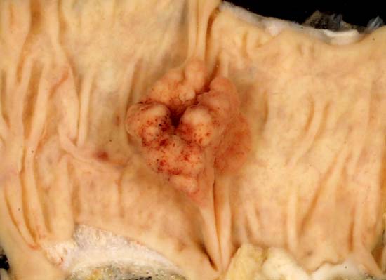
This 2.5-centimeter adenomatous polyp was serendipitously found in a right colectomy specimen. The colon was removed to bypass an extrinsic obstruction caused by metastatic ovarian cancer. In the picture above, the long axis of the opened bowel is horizontal, and the mucosa is viewed en face.
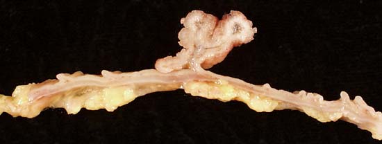
The image above shows a longitudinal section of the same polyp. The normal anatomic layers of the colon are clearly apparent, and it can be seen that the polyp is restricted to the mucosa.
Both photos were shot with an Olympus D-600L digital camera mounted on a copystand illuminated by 4 photofloods. In the top picture, the specimen was submerged in water, while the specimen in the bottom picture was shot dry.
Photograph by Ed Uthman, MD. Public domain. Posted 15 Jan 99
[Back to image table of contents]
[To Ed Uthman's Home Site]
for more original
resources in pathology and laboratory medicine