Hepatocellular carcinoma
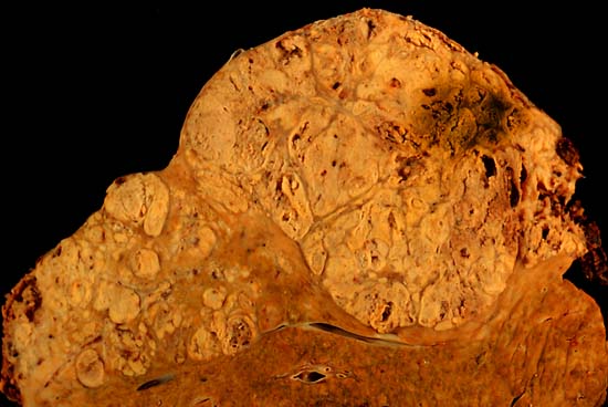
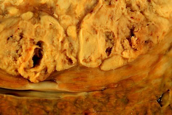
This specimen is from a 50ish woman who presented to the hospital with abdominal pain and ascites. The radiologist recovered what appeared to be whole blood on paracentesis. Cytological exam of the bloody fluid showed no evidence of malignancy. Liver function tests were abnormal, and serologic tests were positive for antibody to hepatitis C. The patient deteriorated rapidly and died within a few days. The autopsy showed this hepatocellular carcinoma occupying much of the volume of a cirrhotic liver. Furthermore, the tumor had invaded the diaphragm and ruptured into the peritoneal cavity, causing the bloody ascites.
The photo above shows a view of a longitudinal slice taken through the full length of the liver. The closer view, below, shows tumor at the top, cirrhotic liver at the bottom, and a fibrous reaction in between. Hepatocellular carcinomas can have a variety of gross patterns, including multinodular/multifocal, such as this one.
The photos were shot with a Minolta X-370 with 100mm bellows lens on Kodak Elite ISO 100 transparency film. The specimen was sliced fresh and fixed in formalin overnight, then briefly immersed in 70% alcohol to retrieve some of the native color and dull the surface reflections.
Photograph by Ed Uthman, MD. Public domain. Posted 23 Sep 00
[Back to image table of contents]
[To Ed Uthman's Home Site]
for more original
resources in pathology and laboratory medicine