Ovarian surface papillary serous tumor
of low malignant
potential
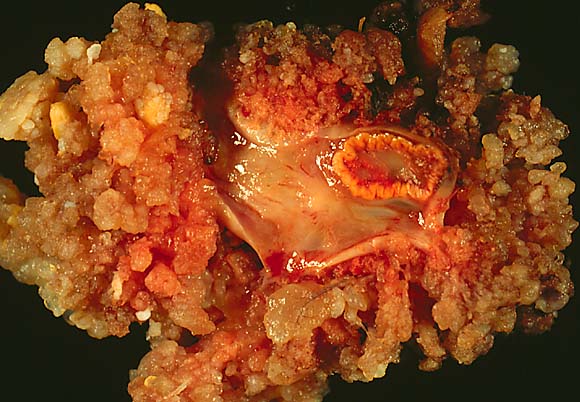
This surface papillary serous tumor of low malignant potential in an oöphorectomy specimen is shot with two techniques. In each photo the ovary is in the center and sports a well-developed corpus luteum in the upper right. The papillary tumor completely surrounds the ovary. This was one of a set of bilateral tumors. The patient had multiple noninvasive peritoneal implants. The image above is shot with conventional copy stand technique and gives maximum resolution.
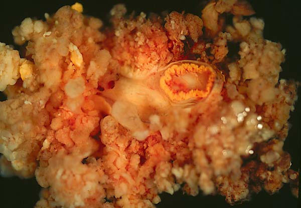
The photo above shows the specimen submerged in water. It is not as sharp or detailed but somehow looks more "biological."
Photograph by Ed Uthman, MD. Public domain. Posted 3 Jan 99
[Back to image table of contents]
[To Ed Uthman's Home Site]
for more original
resources in pathology and laboratory medicine