Metastatic Melanoma in Lymph Node
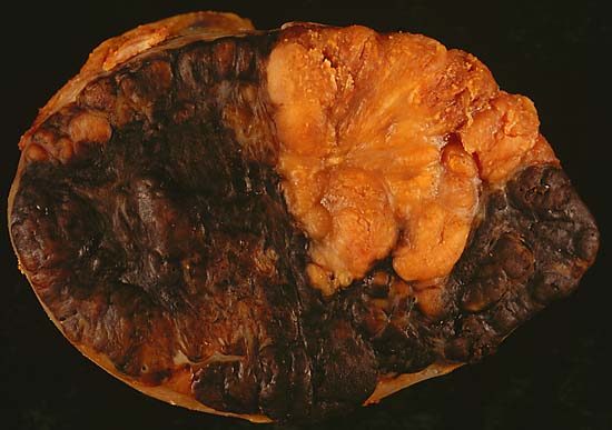
This 6.5-centimeter mass was removed from the axilla of a middle-aged woman, who was a longtime resident of a developmental center for the severely disabled. She was unable to give any history, but there was no medical record of a primary melanoma, and we were unable to find any biopsy scars at the time of surgery. The dark brown color of the cut surface provided a clue to the diagnosis, an unusual finding in metastatic melanomas, which are more typically amelanotic (like the upper right segment of this specimen). Pigment was easily demonstrated in the tumor cells at frozen section, allowing a definitive diagnosis intraoperatively.
Photograph by Ed Uthman, MD. Public domain. Posted 30 May 99
[Back to image table of contents]
[To Ed Uthman's Home Site]
for more original
resources in pathology and laboratory medicine