Bilateral ovarian serous carcinomas
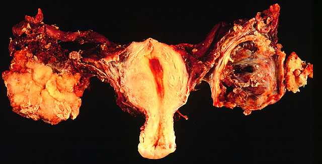
In this TAH-BSO specimen, the right ovary (on the left of the image) has been replaced by a solid serous carcinoma. The contralateral ovarian tumor is grossly cystic and could be termed a "cystadenocarcinoma." The patient had omental metastases and positive peritoneal fluid cytology. This cancer, which was discovered at exploratory laparotomy, apparently developed very rapidly; the patient had a normal pelvic ultrasound exam only 2 months before.

Both photomicrographs are from the solid/papillary right ovarian tumor. As shown in the photo above, much of the tumor had a papillary pattern with exuberant epithelial proliferation but no obvious stromal invasion. Other areas, such as the one depicted in the photo below, show extensive stromal invasion, the criterion upon which rests the diagnosis of frank malignancy.

Photograph by Ed Uthman, MD. Public domain. Posted 2 Jan 99 (updated 6 Feb 99)
[Back to image table of contents]
[To Ed Uthman's Home Site]
for more original
resources in pathology and laboratory medicine