Tubulovillous Polyp
of the Colon
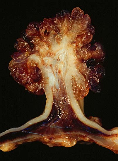
This polyp was removed by segmental sigmoid colon resection from an 18-year-old man who presented with a two-year history of diarrhea. Endoscopically there was a red-hot pancolitis, which histologically showed a brisk neutrophilic cryptitis with numerous crypt abscesses. Convincing features of chronicity were elusive, a few basilar plasma cells notwithstanding. The head of the polyp showed adenomatous architecture with both tubular and glandular elements. The stalk contained a pipechase full of large blood vessels, attesting to the wisdom of not snaring this 2-cm polyp endoscopically.
After the initial colonoscopy and biopsy, the presumptive diagnosis of chronic ulcerative colitis (CUC) with dysplasia-associated lesion or mass (DALM) was tendered, and the patient was started on anti-colitic therapy. He responded quickly--a little too quickly in the eyes of the clinical team. Sure enough, within a few days, the segmental resection showed complete resolution of the inflammation with no evidence of chronic mucosal damage. At this point, what appeared to be ulcerative colitis with DALM, is now putatively a coincidence of two independent lesions, a rare tubulovillous polyp in a teenager, and acute self-limited colitis. Needless to say, the patient will be followed closely to make sure he does not have CUC.
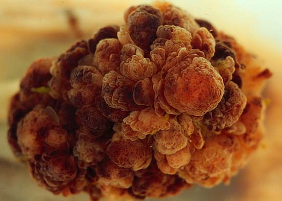
The photograph above shows the polyp viewed "down" from "above" its head, looking down toward the out-of-focus native mucosa from which it arises. This polyp is ususually dark because of the bleeding into the mucosa and submucosa caused by the abortive attempts at endoscopic removal.
The polyp is shown in longitudinal section in the photo below. Resembling congealed beef buillon, the hemorrhagic submucosa of the stalk and colon wall proper is set off strikingly from the pale ribbon of mucosa that covers it.
Both photos were taken with a Minolta X-370 with 100mm Rokkor bellows lens, illuminated by 4 photofloods, on Kodak Elite ISO 100 daylight slide film, with 80B blue filter to compensate for tungsten color temperature.
As revealed by the complete absence of highlights, the top photo was shot under water. I think this technique is especially useful for delicate villous structures that would collapse under their own weight if not suspended in a liquid medium of like density. Submersion also softens and thus de-emphasizes distracting backgrounds, like the normal colonic mucosa deep to the polyp head.
Photograph by Ed Uthman, MD. Public domain. Posted 28 May 00
[Back to image table of contents]
[To Ed Uthman's Home Site]
for more original
resources in pathology and laboratory medicine