Villous adenoma
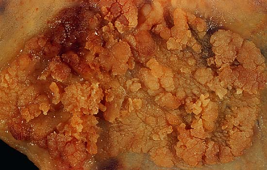
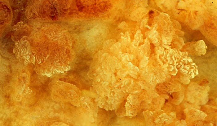
This large (6.5 cm) villous adenoma of the sigmoid colon had no invasive component, so the patient's prognosis after excision is excellent. The photo above is shot using conventional copy stand techniques and shows what this tumor looked like to the pathologist on the gross board, and to the gastroenterologist through the endoscope. The photos below show the specimen as immersed in tapwater. The buoyancy of the delicate villous structures causes them to stand up and separate, showing the complex coral-like filigree produced by this type of growth pattern.

The image below is not a close-up as such; it is simply a high-resolution scan of the transparency reproduced immediately above. Scanned at near the maximum resolution of the Polaroid SprintScan scanner, this image shows the detail that Ektachrome Elite is capable of.
All photos were shot with a Minolta X-370 with bellows lens, on Ektachrome Elite 100 film, daylight type, through a blue filter to correct for Photoflood ilumination. Exposure was f/8 at something like 1/2 or 1 second.
Photograph by Ed Uthman, MD. Public domain. Posted 27 May 01
[Back to image table of contents]
[To Ed Uthman's Home Site]
for more original
resources in pathology and laboratory medicine