
MICROWAVE LABORATORY
DEPARTMENT OF PHYSICS, IIT MADRAS, CHENNAI - 600 036
Dr. V. Subramanian Email: manianvs(at)iitm.ac.in
Associate Professor
SCANNING NEAR FIELD MICROWAVE MICROSCOPY
Imaging is a versatile tool to understand the spatial variation of any physical parameter such as temperature, reflectivity etc. of any object or scene. In a far-field methodology, we use elements that satisfy Fraunhofer diffraction and the spatial resolution of such image is limited by aperture and wavelength. Therefore to develop a microscope with micrometer resolution, one has to use wavelength of the order of 500 nm or less.
In the near-field imaging, the evanescent wave from an
illuminated tiny aperture can be used to image the object. In this method,
the diffraction limited case is not obeyed and the spatial resolution can be
obtained as small as 10-5 times of the wavelength. The
parameters that decide the spatial resolution are:
a. The distance between the aperture and the object
b. The size and shape of the aperture and
c. The electric or magnetic field pattern of the evanescent wave.
The microscope that operates at microwave frequencies (with wavelengths of the order of few centimeters) can give a spatial resolution of the order of 1 micrometer (similar to the Scanning Near field Optical Microscope where sub-nm resolution is achieved) and even less than that.
Our microwave laboratory has now the capability of spatial resolution of the order of 1 microns.
Applications?
Microwave has the capacity of penetrating through thin films, polymer sheets etc. and therefore can be used to probe the surface of the metals covered by such materials.
Can probe the dielectric as well as magnetic inhomogeneity on the surface.
Surface scanning of semiconductor wafers for conductivity and carrier lifetime.
Grain boundary studies in polycrystalline materials.
Microwave Hall effect mapping of semiconductors.
And so on........
Some of our theoretical and experimental results:
  |
 
Experimental result obtained for cavity method of SNMM for Air-metal interface |
|
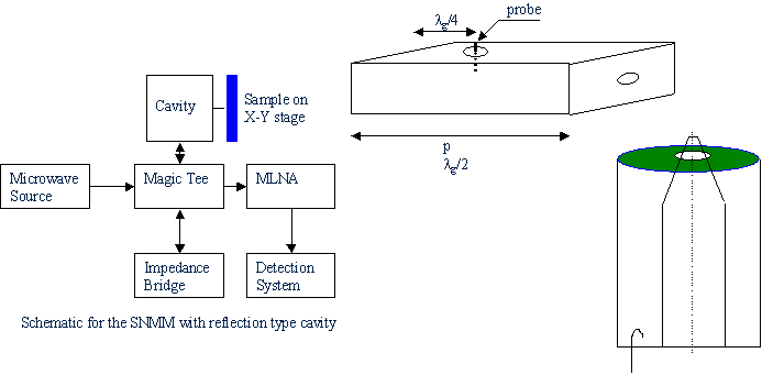
Our experimental arrangement with coaxial probe and the obtained result: metal plate with hole at the center. The image is 1/4th of the hole. |
|
Recent image obtained on glass-metal-glass interface - We have achieved a spatial resolution around 1 microns. |
List of publications related to SNMM: Yet to open the account