Baby Explorer™
by
CEDARA Co.
Download Free Baby Explorer Trial Version
3D Fetal Imaging The Baby Explorer™ software-only solution allows you to easily and quickly generate fetal images months before the baby is born. With a minimal amount of training, the Technologist can quickly generate an image of the baby’s facial features and print it to a Microsoft Windows printer. The image can also be saved as a JPEG, BMP, or AVI file.
Key Benefits
- Works with any ultrasound system
- Easy acquisition controls
- 3D segmentation and clipping tools
- Built-in animation and multimedia export capabilities
- Prints to standard printers in variety of formats
Baby Explorer software is a cost effective, software-only solution that enables the technologist to quickly and easily generate 3D fetal views. With its sophisticated volume rendering capabilities, the Baby Explorer software enables the anatomical features of the baby to be seen. Ultrasound 3D neonatal studies are used to ensure the proper growth of the fetus and to investigate cleft palette and other gross abnormalities. The Baby Explorer software runs on standard PC hardware and acquires images from any ultrasound scanner.
Key Features
- Works with any ultrasound system
- Easy acquisition controls
- Multiplanar Reformatting (MPR)
- Opacity rendering
- Gradient shaded rendering
- 3D tissue segmentation and clipping tools
- Animation and multimedia export
- Prints to standard printers
- HTML reporting
Data Management Patient
folder data management (i.e., start, open and delete)
Acquisition
- Captures ultrasound images directly from conventional ultrasound systems
- Allows video setting adjustments
- Able to acquire 2D sweeps in either linear or fan motion
- Displays live mode allows previews of the selected sweep
- Able to modify ROI after an acquisition is performed
- Allows the display of the current sweep in the 3D window
3D Image Review
- Four viewports to display the sweep: 3D surface rendered, axial, coronal, and sagittal
- 3D image manipulation tools (e.g., window, zoom, pan)
- Advanced smoothing algorithm to smooth the surface of the volume on large data sets
- Able to adjust the opacity to cut the noise levels and improve the quality of the rendered volume
- Able to create VOI from clippers to explore the volume
- Front Cut Clippers — allows exposure of the structures inside the volume
- Bubble Clippers — allows display of the volume contained in the circle clipper
- Segmentation — able to remove noise in the volume using a scalpel tool
- Able to adjust the geometry (i.e., change scan types, reverse slide order, change width of the volume)
HTML Reporting
- Able to add text comments to the study as an HTML file
- Able to include the 2D cine of the fetus as an AVI file and the 3D volume rendered image of the fetus as a JPEG image
Printing and Multimedia
- Prints to standard printers and exports to standard image formats (i.e., BMP and JPG)
- Able to export the animated 3D volume to AVI format
- Able to print and export multiplanar images
Minimum System Requirements
- Microsoft® Windows® XP, 2000, ME, 98
- Intel® Pentium® III 800Mhz
- 128 Megabytes RAM, 20-Gigabyte hard disk
- 32-bit color display video card and video capture card
Click here to see more pictures made by Baby Explorer - Gallery 2
|
|
|
|
|
|
|
|
|
|
|
|


Click here to see more pictures made by Baby Explorer - Gallery 2
 |
 |
 |
 |
 |
 |
|
Blood vaseles of folicles beffore ovulation |
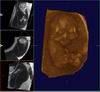
18 weeks Baby Explorer |
12 weeks Baby Explorer |
12 weeks Baby Explorer |
11 weeks (male) Baby Explorer |
11 weeks Baby Explorer |
 |
 |
 |
 |
 |
|
|
29 weeks Baby Explorer |
29 weeks Baby Explorer |
29 weeks Baby Explorer |
12 weeks |
12 weeks
movie |
(Click over link if you wish to see some short clips (made by Baby Explorer ™)
-
Corpus Luteum Cyst scanned by free hand technique (Baby Explorer reconstruction)*
-
Embryo at 9 weeks scanned by free hand technique (Baby Explorer reconstruction)
-
Same multilocular ovarian tumor scanned by free hand technique (Baby Explorer reconstruction)**
-
Missed abortion (10 weeks) scanned by free hand technique (Baby Explorer reconstruction)***
-
Same missed abortion (10 weeks) scaned by 3D live technique***
-
Fetus at 12 weeks scanned by free hand technique (Baby Explorer reconstruction)
-
Fetus at 12 weeks scanned by free hand technique (Baby Explorer reconstruction)
-
Fetus at 15 weeks, legs, abdomen and pelvis (Baby Explorer software reconstruction)
-
3D reconstruction of ovarian cancer (Baby Explorer software reconstruction)
-
3D reconstruction of ovarian cancer (Baby Explorer software reconstruction)
-
3D reconstruction - Blood vessels of pregnant uterus (7 weeks)
-
Fetus at 12 weeks scanned by free hand technique (Baby Explorer)****
-
Same fetus 12 weeks scanned by 3D live technique (Voluson)****
-
3D reconstruction - fetus at 14 weeks - Baby Explorer volume 1 and volume 2
- Blood vaseles of folicles beffore ovulation
- 3D reconstruction of fetus at 11 weeks (Baby Explorer software reconstruction)
- 3D reconstruction of fetus at 12 weeks (Baby Explorer software reconstruction)
- 3D reconstruction of fetus at 18 weeks (Baby Explorer software reconstruction)
If you need help working with Baby Explorer ™ please do not hesitate to contact me.
If you need help creating hybrid system 2D ultrasound-software-PC please do not hesitate to contact me.
Transformation of 2D ultrasound machine to 3D (even 4D) system is easy and fast work.
Copyright © 2008. Design by Ivan Bubanovic