Paul Murata
Osteochondritis Dissecans of the Knee
Introduction:
In this paper I will identify and discuss the knee disorder of Ostechondritis Dissecans (OCD) . I will start with the identification of OCD and then move on the injury evaluation of the injury. I will concentrate on OCD of the lateral aspect of the medial condyle. I will follow the evaluation by addressing any adjacent joint considerations and then end with my conclusions.
Identification:
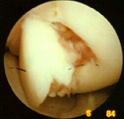
Osteochondritis Dissecans is a defect in the subchondral region with partial or complete separation of the cartilage and subchondral bone from an articular surface (2,3,7). OCD may affect many joints such as the hip, knee, ankle, elbow or shoulder (2,11,12). OCD of the knee is most often seen on the postero-lateral aspect of the medial femoral condyle ( 80% - 85% of cases) (figure 1 & 2) (2,11), and is usually unilateral (74%) (figure 3) (7,12). The lateral femoral condyle on the patella may also be affected, but these condition are very rare (figure 1b&c ) (2,3,4,5,7,10,12). OCD is more common in males (ratio male:female , 2:1 – 3:1) and usually occurs between the ages of ten and fifty, with the average age being 18 (2,3,7,12). The peak incidence of OCD occurs at puberty (11).
Evaluation:
Injury mechanism
There is no clear cause of OCD, however it has been thought to be caused by trauma, ischaemia, or genetic predisposition (2,3,7,8,12).
Trauma
May be due to a twisting injury to the knee, or a patellar dislocation (3). Chronic, repetitive knee trauma may also play a role in the onset of OCD. This is when the repetitive overloading causes fragmentation and separation of bony fragments (2,3,7,12). This recurrent trauma may also cause a loss of circulation to the area (8).
Ischaemia
Sometimes the interruption of blood supply to the region will cause the onset of OCD (3,6,). Cartilage, nourished by synovial fluid, may begin to soften and degenerate with repetitive trauma. (2). In the maturing child, the presence of an epiphyseal plate will affect blood supply to the immature bone ends (11).
Genetic Predisposition
It is possible that one can be predisposed to OCD depending if there is a family history of the disorder (2,3,10,12)
Inspection-Observation
The complaint of pain from the athlete will usually be vague. The athlete may complain of aching with and after exercise combined with joint effusion (2,3,7,9,12). There also may be complaints of the knee locking, giving way, or crepitation during knee flexion and extension (2,3,7,9,10,12). With the lateral aspect of the medial condyle affected with OCD, the athlete will complain of pain when cutting or internally rotating their tibia (2,3,7,). As a result of this pain, athletes may walk with the affected leg externally rotated (2). Muscle atrophy and mild joint effusion may also be observed (3,9,12,13). Knee abnormalities such as genu valgum and genu varum may also be associated with OCD (2).
Pain and Palpation
Pain is reproduced with deep palpation to the lateral aspect of the medial femoral condyle (9,12). The athlete may have a palpable loose body, but this is less common in younger athletes(3). With forcible compression to the affected side of the knee, joint may elicit crepitation during knee flexion and extension.
Range of Motion
Typically, the knee maintains full range of motion (0 – 135 degrees flexion, 0 – negative 5 degrees extension), unless a loose body causes mechanical locking (2,3,12). Loss of terminal flexion due to joint effusion may be noticed in some cases (3). Passive range of motion with an internally rotated knee may cause pain (7).
Special Tests
The widely used clinical test for OCD is the Wilson Test. With the athlete seated with the knee flexed 90 degrees, the athlete is asked to slowly extend the knee while the tibia is internally rotated(figure 4). The athlete is then asked to stop extending when pain is experienced and hold that position. If OCD is present, the pain will be relieved by the athlete externally rotating their tibia (1,2,3). Athletes with OCD usually experience pain at 30 degrees flexion (1,2,3).
Manual Muscle Tests
Manual muscle tests may not reveal a great deal of information when assessing OCD. Pain may be present during knee extension, but not if the tibia is externally rotated (2,7,9). A difference in quadracep atrophy and strength may be noticed, but only if one knee has OCD (2,3,7,9). Crepitation may be noticed while performing manual muscle tests on the knee (7).
Neurovascular:
This subject is not applicable.
Functional:
Functionally, the athlete will have pain during cutting or twisting actions of the lower leg. In order to avoid impinging the lesion on the medial femoral condyle, the athlete may walk with their leg externally rotated (2,3,7).
Diagnostic:
An athletic trainer can suspect OCD through clinical evaluation, but OCD can only be diagnosed by a doctor and confirmed by radiographic (figure 3) or MRI (figure 5a,b&c) findings (2, 7,11). Radiographs will show OCD as a fracture to the condyles. MRI findings may be used to determine whether the fragment is detached or not (7). There are four classifications of OCD (3,7) and they are:
I) Depressed osteochondral fracture;
II) Osteochondral fragment attached by an osseous bridge
III) Detached non-displaced fragment;
IV) Displaced Fragment;
Adjacent Joint Considerations:
Although OCD is usually unilateral (74%) (3,7,12), adjacent joint considerations should always be addressed. If the OCD is due to a genetic prediposiosion then the athlete may suffer from OCD in a multiple number of different joints: hip, ankle, elbow or shoulder (2,11,12).
Conclusion
:
Due to the mystery surrounding the exact cause of OCD, athletic trainers may find it difficult to prevent the disorder. However, through careful and attentive evaluation, an athletic trainer can help the physician make positive diagnoses. By performing proper and precise tests OCD can be identified at an early stage. Although OCD is relatively uncommon, it should be considered when dealing with young athletes and chronic knee problems (2,4,11).
