
FIG 1. Large intra-abdominal tumor arising from left ovary.
Abdominal masses in children are uncommon. Ovarian neoplasms are uncommon in young children and adolescents, and typically are benign. Small cell carcinoma of the ovary is an extremely rare condition, with a very poor prognosis. We report the case of a 15-year-old female who presented to the emergency department with abdominal distention, abnormal vaginal bleeding, and constipation secondary to a large intra-abdominal mass, ultimately diagnosed as ovarian small cell carcinoma.
Abdominal masses are uncommon in young children and adolescents. Although rare, ovarian neoplasms must be considered in the differential diagnosis of a painless abdominal mass in young females. Small cell carcinomas are extremely rare neoplasms of the ovary, which act in an aggressive manner. When the diagnosis of small cell carcinoma is made, the prognosis is very poor. Treatment generally is ineffective, and only a few patients have survived more than 2 years. This case report describes a patient who presented with a large, painless, abdominal mass, which was eventually diagnosed as ovarian small cell carcinoma.
Her medical history was significant for the following: being born at 29 weeks' gestational age, scoliosis, and an anxiety disorder for which she took sertraline {ZoloftTM }. She experienced menarche at age 12. Her menstrual periods were described as irregular. She denied any prior sexual intercourse, and there was no family history of gynecologic cancer.
FIG 1. Large intra-abdominal tumor arising from left ovary.
The physical examination revealed a well-developed female in no distress. Vital signs were temperature 98.8"F, pulse I00 bpm, respirations l 8/min. and blood pressure of 100/70 mmHg. She had a distended abdomen with a firm, non-pulsatile, palpable mass beginning in her pelvis and extending to her umbilicus. There was mild periumbilical tenderness, but no guarding or rebound. There was no hepatomegaly or splenomegaly. There were good bowel sounds, and the abdomen was non-tympanitic. Rectal examination was negative for occult blood, and there were no masses. There was no inguinal adenopathy. She had no costovertebral angle tenderness, no lower extremity edema, and good distal pulses.
A urine pregnancy test was negative. Initial laboratory results, including complete blood count, electrolytes, blood urea nitrogen, creatinine, and glucose, showed normal values. A calcium level was not obtained at this point. An abdominal computed tomographic (CT) scan was obtained to further define the mass. The CT scan showed a 12 x 13 x 16 cm multiloculated cystic mass, most likely of ovarian origin (Fig. I). Marked ascites and bilateral pleural effusions were also evident (Figs. 2 and 3). Gynecologic consultation was obtained in the ED, and arrangements were made for outpatient referral to a gynecologic oncologist. A complete gynecologic examination was deferred at this point.

FIG 2. Marked ascites.
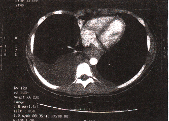
FIG 3. Bilateral pleural effusions.
Six days after presentation to the ED, the patient underwent an exploratory laparotomy. A 17 x 15 x 10 cm, multiloculated smooth-surfaced mass was found and removed, and a left salpingo-oophorectomy was performed. A subclavian mediport was placed in preparation for chemotherapy.
On gross pathologic examination, sections through the ovary showed a "predominately solid to focally cystic variegated, tan to pink, focally necrotic, focally hemorrhagic tumor." Final microscopic specimens determined the tumor to be a small cell carcinoma, hypercalcemic type. There was some debate as to whether pelvic lymph node sampling was positive for small cell carcinoma versus atypical mesothelial hyperplasia. The tumor was classified as stage T1CN1.
The patient has since undergone two cycles of chemotherapy with etoposide, ifosfamide, and cisplatin. Her initial CA125 level decreased to 13.1 from 208.9 after the first cycle. A serum calcium level obtained prior to initiation of chemotherapy was within normal limits at 9.6. A routine CT scan of her chest 3 weeks postoperatively showed two right-sided, pleural-based metastases and an incidental occlusion of her right internal jugular vein. Heparinization was instituted, and warfarin was subsequently added. She is currently awaiting a stem cell rescue following pheresis.
The incidence of ovarian cancer for women less than 30 years old is lower than 3.0 per l00,000 population; for women over 70 years, the incidence rises to 54.4 per 100,000 (1,2). Ovarian neoplasms are rare in children, constituting only 1% of all pediatric cancers (3,4). Most ovarian masses in infants and young children are benign (80%); the most common type is the teratoma (5).
Whereas epithelial tumors are most common in adults, tumors of germ cell and sex cord stromal origin predominate in children (2, 3, 6-14). Germ cell tumors include benign cystic and malignant teratomas, dysgerminomas, yolk sac tumors, embryonal carcinoma, and choriocarcinoma (12, l5). The low incidence of epithelial neoplasms in childhood is felt to be due to a lack of gonadotropic hormonal stimulation (4). Other rare tumors of the ovary found in children include fibrosarcoma, cystadenomas, cystadenocarcinomas, granulosa cell carcinomas, mesonephric carcinoma, lymphangioma, arrhenoblastoma, and metastatic intestinal lymphomas and adenocarcinomas (3, 5, 7-9, 12).
Small cell carcinoma of the ovary is a very rare malignancy, first reported in 1982 by Dickersin et al. (16). Since 1982, 166 cases have been described (17). These tumors are very aggressive and affect a younger age group than other ovarian malignancies (l7-l9).
They often show widespread intra-abdominal metastases (20). Hypercalcemia is seen concomitantly in approximately two thirds of cases (20), and calcium levels often return to normal after treatment is initiated, or increase during relapse (18,19). Elevated calcium levels are thought to arise from ectopic tumor production rather than by bony metastases (18).
Clinically, patients with ovarian cancer typically present with abdominal pain, abdominal distention, or pelvic pressure (1). Torsion may occur as the tumor enlarges into the abdomen and elongates the pedicle or ovarian ligament (5,9). Amenorrhea, anorexia, vomiting, infertility, and weight loss have also been described with small cell carcinoma (18, 21). When hypercalcemia is present, the symptoms of nausea and vomiting may be attributable to the high calcium levels and not to the tumor per se (21). Ultrasonography or computed tomography (CT) is helpful in delineating the size and complexity of these tumors.
Metastases and associated pleural fluid collections are readily detected by CT. The histologic diagnosis is often difficult and electron microscopy is often needed to make the final diagnosis (18). Originally thought to arise from an epithelial cell line (16,22), there is debate over the histogenesis of these tumors. A germ cell theory has been favored by Ulbright et al. (23), whereas Eichorn et al. (24) favor a sex cord origin.
Survival rates for all malignant ovarian neoplasms have been poor (7). Ovarian small cell carcinoma has a particularly dismal prognosis. Despite various treatment modalities including resection, radiation therapy, and intensive chemotherapy, mortality for small cell carcinoma remains high, with the average life expectancy being 18 months (17,18,21). Various chemotherapy regimens have been described, including combinations of vincristine, cisplatin, actinomycin D, cyclophosphamide, bleomycin, dacarbazine, vinblastine, etoposide, and doxorubicin; these regimens have provided minor or inconclusive benefit (18,19). There are only four cases of long-term surviving patients (16, 19-21).
From the Department of Emergency Medicine, Albany Medical Center, Albany, New York.
Address for reprints:
Peter C Ferrera, MD
Department of Emergency Medicine Mailcode-139
Albany Medical Center
43 New Scotland Avenue
Albany, NY 12208
Key Words: Abdominal mass, ovarian carcinoma, neoplasm, small cell carcinoma