![]()

The Anatomy of the Immune System
Dr K. West
Aims and Objectives
By the end of the module you should be able to:- understand the concept of bone marrow stem cells giving rise to the cells of the immune system
- describe the anatomical location of the thymus gland
- describe the structure of the thymus gland
- cortex
- medulla
- Hassall's corpuscles
- describe the cellular composition of the thymus gland
- describe the passage of cells through the thymus gland during the process of T cell maturation
- understand the changes that occur in the thymus gland with age
- describe the anatomy of the lymphatic system including the general structure of lymph nodes
- describe the cellular composition of lymphoid follicles, paracortex and medulla
- understand the role of high endothelial venules in lymphocyte traffic
- describe the passage of lymph through lymph nodes and understand how this facilitates the immune response to antigens
- describe the anatomical location of the spleen
- describe the splenic blood supply
- describe the cellular composition of the red and white pulp
- understand the contribution of the spleen to the immune response
- predict the possible effects of splenectomy
- describe the common sites and the cellular constituents of mucosa associated lymphoid tissue
- describe the process and purpose of lymphocyte recirculation
- cortex
Introduction
The immune system operates throughout the body. There are, however, certain sites where the cells of the immune system are organised into specific structures. These are classified as central lymphoid tissue (bone marrow, thymus) and peripheral lymphoid tissue (lymph nodes, spleen, mucosa-associated lymphoid tissue):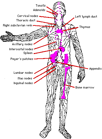
1. Bone marrow
All the cells of the immune system are derived from stem cells in the bone marrow. The bone marrow is the site of origin of red blood cells, white cells (including lymphocytes and macrophages) and platelets. The cells of the immune system are considered in detail elsewhere.2. Thymus
In the thymus gland lymphoid cells undergo a process of maturation and education prior to release into the circulation. This process allows T cells to develop the important attribute known as self tolerance.a) Anatomy: The thymus
gland is found in the thorax in the anterior mediastinum. It gradually
enlarges during childhood but after puberty it undergoes a process of
involution resulting in a reduction in the functioning mass of the gland.
It continues to function throughout life, however.
b) Histology: The thymus gland is
arranged into an outer, more cellular, cortex and an inner, less cellular,
medulla. Immature lymphoid cells enter the cortex proliferate, mature
and pass on to the medulla. From the medulla mature T lymphocytes enter
the circulation.
 |
The following cell types are present:
- lymphoid cells
- epithelial cells
- macrophages
- other supporting cells
Thymic epithelial cells have different appearances in different locations within the gland. They form a continuous sub-capsular layer and a network in the cortex and medulla. Deep in the medulla they are also aggregated into Hassall's corpuscles.
3. Lymph nodes
Lymph nodes are small bean shaped structures lying along the course of lymphatics. They are aggregated in particular sites such as the neck, axillae, groins and para-aortic region. Knowledge of the sites of lymph nodes is important in physical examination of patients.Lymph nodes have two main functions:
- phagocytic cells act as filters for particulate matter and micro-organisms
- antigen is presented to the immune system
a) Structure
Lymph nodes have a fibrous capsule from which trabeculae extend towards the centre thus forming a framework:
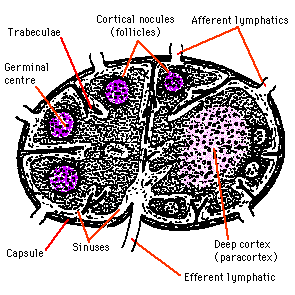 |
The node is made up of three components:
- lymphatic sinuses
- blood vessels
- parenchyma (cortex, paracortex, medulla)
b) Cortex
B cells: These enter the lymph node via HEVs and pass to the follicles. If activated by antigenic stimulation they proliferate and remain in the node. Unstimulated B cells, however, pass out rapidly from the node to return to the general circulation. Activated B cells within the lymphoid follicles are known as follicle centre cells. The pale staining central area of a secondary follicle is known as a germinal centre and this is surrounded by a mantle zone consisting of small, naive B cells and a few T cells.The follicle centre cells within the germinal centres consist of cells with cleaved nuclei (centrocytes) and cells with larger more open nuclei and several nucleoli (centroblasts).
Stimulated mature B cells responding to antigen change into centrocytes and then centroblasts. The centroblasts leave the follicle and pass to the paracortex and medullary sinuses, where they become immunoblasts. The immunoblasts divide to give rise to plasma cells or memory B cells which are ready for their next encounter with specific antigen.
Accessory cells: Lymphocytes alone are not to make an effective immune response. They are assisted by so-called accessory cells. These may be grouped as follows:
- sinus macrophages (highly phagocytic)
- tingible body macrophages (ingest cellular debris in germinal centres)
- marginal zone macrophages (found beneath the subcapsular sinus)
- follicular dendritic cells
c) Paracortex
The paracortex contains lymphocytes and accessory cells along with supporting cells and it is the predominant site for T lymphocytes within the lymph node.T cells: The various types of T cell enter the node from the blood via the HEVs. When activated they form lymphoblasts which divide to produce a clone of T cells responding to a specific antigen. Activated T cells then pass into the circulation to reach peripheral sites.
Accessory cells: Interdigitating cells are numerous in the paracortex and they act as antigen presenting cells.
d) Medulla
The medulla comprises:- large blood vessels
- medullary cords
- medullary sinuses
e) Passage of lymph
Lymph passes into the node through the afferent lymphatic into the marginal sinus, though the cortical sinuses to reach the medullary sinuses before leaving via the efferent lymphatic. Particulate matter in the lymph is removed by macrophages. Antigens are taken up by antigen presenting cells and these facilitate the specific immune response. Less than 10% of lymphocytes enter the node in the lymph, the large majority entering from the blood via the HEVs.4) Spleen
The spleen is located in the upper left quadrant of the abdomen. It has two main functions acting as part of the immune system and as a filter.a) Structure
The spleen has a thin connective tissue capsule from
which short septa extend inwards. These septa are, in turn, connected
to a complex reticulin framework.
There are two distinct components of the spleen, the red pulp and the
white pulp. The red pulp consists of large numbers of sinuses and sinusoids
filled with blood and is responsible for the filtration function of the
spleen. The white pulp consists of aggregates of lymphoid tissue and is
responsible for the immunological function of the spleen:
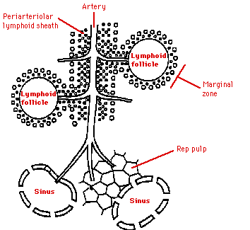 |
b) Red pulp
There is a complex system of blood vessels within the red pulp arranged to facilitate removal of old or damaged red blood cells from the circulation. A small proportion of the splenic blood flow passes through more rapidly without undergoing this process of filtration.c) White pulp
The white pulp contains T cells, B cells and accessory cells. There are many similarities with lymph node structure. The purpose of the white pulp is to mount an immunological response to antigens within the blood. The white pulp is present in the form of a periarteriolar lymphoid sheath. This sheath contains B cell follicles and T cells. At the edge of the T zone is a region known as the marginal zone where larger lymphocytes and antigen presenting dendritic cells are located.5) Mucosa-associated lymphoid tissue (MALT)
In addition to the lymphoid tissue concentrated within the lymph nodes and spleen, lymphoid tissue is also found at other sites, most notably the gastrointestinal tract, respiratory tract and urogenital tract.Gut associated lymphoid tissue (GALT)
This comprises:- tonsils, adenoids (Waldeyer's ring)
- Peyer's patches
- lymphoid aggregates in the appendix and large intestine
- lymphoid tissue accumulating with age in the stomach
- small lymphoid aggregates in the oesophagus
- diffusely distributed lymphoid cells and plasma cells in the lamina propria of the gut
Peyer's Patches
These are quite large aggregates of lymphoid tissue found in the small intestine. The overlying 'dome' epithelium contains large numbers of intraepithelial lymphocytes. Some of the epithelial cells have complex microfolds in their surfaces. They are known as M cells and are believed to be important in the transfer of antigen from the gut lumen to Peyer's Patches. Peyer's Patches facilitate the generation of an immune response within the mucosa. B cell precursors and memory cells are stimulated by antigen in Peyer's Patches. Cells pass to the mesenteric lymph nodes where the immune response is amplified. Activated lymphocytes pass into the blood stream via the thoracic duct. These cells then home in the gut and carry out their final effector functions. HEVs are not present in Peyer's Patches and the mechanism by which cells home in on mucosal sites is unknown. Cell surface molecules known as addressins may have a role.6) Lymphocyte recirculation
Lymphocytes and some mononuclear phagocytes can recirculate between lymphoid and non-lymphoid tissues. This helps in allowing lymphocytes to be exposed to the antigens which they recognise and is, therefore, valuable in the distribution of effector cells of the immune response to the sites where they are needed:
 |
The recirculation is a complex process depending on interactions between the cells of the immune response and other cell types such as endothelial cells. virgin lymphocytes move from the primary to secondary lymphoid tissue via the blood activated lymphocytes move from the spleen, lymph nodes and MALT into the blood and thence to other lymphoid and non-lymphoid tissues antigen presenting cells such as macrophages and dendritic cells may carry antigen back to lymphoid tissues from the periphery. The complex patterns of recircultion depend on the state of activation of the lymphocytes, the adhesion molecules expressed by endothelial cells and the presence of chemotactic molecules which selectively attract particular populations of lymphocytes or macrophages.
 |
![]()
If this information was useful, read this
Search for more information on this topic
Contents | Previous Page | Next Page
![]()
Webmaster : Mr. Kulachat Sae-Jang
4436692 MTMT/D
Medical Technology, Mahidol University
E-mail address : oadworld@yahoo.com