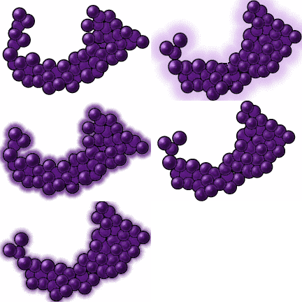
"Staphylococcus bacterial cells are spherical, 0.5-1.5 micrometers in diameter and occuring singly, in pairs and characteristically dividing in more than one plane to form irregular clusters. This genus is non-motile and gram-positive. The cell wall contains two main components; a peptidoglycan and its associated teichoic acids. The peptidoglycan consists of glycan composed of repeating units of Beta-1,4 linked N-acetyl-glucosamine and N-acetyl-muramic aced residues that are linked through N-acetyl-muramyl-L-alanine linkages to peptide subunits. The peptide subunits are cross-linked by penta-peptide bridges containing solely or mainly glycine. These bridges extend from the N-lysine residue of one peptide subunit to the C-terminal D-alanine of a neighboring peptide subunit. Attached to or in close proximity to the peptidoglycan are teichoic acids which, depending on the species, contain either ribitol or glycerol linked to a sugar or an aminosugar."1 "Staphylococcus, genus of round, parasitic bacteria, commonly found in air and water and on the skin and upper part of the human pharynx. These bacteria are known to cause pneumonia and septicemia as well as boils and kidney and wound infections (see Abscess; Carbuncle; Infection). The antibiotic drug penicillin was once effective for the treatment and control of staphylococci, but the increase of resistant strains requires use of other antiobiotic agents such as semi-synthetic penicillins, cephalosporins or vancomycin. Two common species of Staphylococcus include Staphylococcus aureus, which is commonly responsible for skin infections, and Staphylococcus epidermis, which does not normally cause infection. However, either of these bacteria can cause serious infections under the right conditions. S. aureus is found on the skin and in the nostrils of many healthy individuals. These bacteria often give rise to minor superficial diseases, including the formation of pustules or boils in hair follicles. S. aureus infections are characterized by the presence of pus and formation of abscesses. In addition to skin pustules, boils, and carbuncles, S. aureus is responsible for impetigo, infections of wounds and burns (particularly in a hospital environment), breast abscesses, whitlow (inflammation of a finger or toe near the nail), osteomyelitis, bronchopneumonia, septicemia, acute endocarditis, food poisoning, and scalded skin syndrome. Scalded skin syndrome occurs in newborns and is due to infection by toxigenic strains of S. aureus. The toxins cause the skin to exfoliate, which leaves an appearance of having been scalded. S. epidermis does not usually cause infection, occurring universally in a harmless symbiotic relationship (see Symbiosis). It is usually present on most areas of the skin, in the nostrils, mouth, external ear, and urethra. However, S. epidermis can take advantage of a host with a suppressed immune system and can aggravate an existing condition. Following heart surgery, S. epidermis may cause endocarditis. S. epidermis may turn an existing abnormality in the urinary tract into cystitis."2 "Staphylococci cause abscesses, boils, and other infections of the skin, such as impetigo. They can also produce infection in any organ of the body (e.g., staphylococcal pneumonia of the lungs). The most common form of food poisoning is brought on by staphylococcus-contaminated food. The staphylococcus organisms also generate toxins and enzymes that can destroy both red and white blood cells."3 "Staphylococcus aureus: Cells occur singly, in pairs and divide in more than one plane to form irrelgular clusters. Some strains possess a capsule or slime layer. Cell walls contain organic phosphorous, ribitol, glucosamine, muramic acid, blycine, lysine, aspartic acid, serine, glutamic acid, alanine and small amounts of threonine, proline, valine and leucine. These compounds can be accounted for in three of the main components of the wall which are the peptidoglycan, the ribitol teichoic acids and the species-specific precipitinogen Protein A. Cell membranes containe the glycolipids, mono and diglucosyl-diglyceride and the phospholipids, lysyl-phosphatidyl-glycerol, phosphatidyl-glycerol and cardiolipin. The colonies are smooth, low-convex, glistening, butyrous and with an entire edge. Under conditions inhibitory to normal growth, rough and dwarf colony variants may be produced. Colonial pigmentation is extremely variable. Colonies of most strains are orange in color although certain antibiotic-resistant strains and strains from bovine sources are more commonly yellow pigmented. Their growth on agar slants is abundant, they are opaque, smooth, flat, moist and white, yellow or orange in color. All strains are potential pathogens. Under suitable conditions strains produce a variety of enzymes believed by some to play a role in initiating infection; these include hyaluronic acid lyase, fibrinolysin, nucleases and coagulases. Certain antigenic factors are thought to play a part in enabling a strain to set up an infection. The symptoms may be caused by a variety of toxins, the most immportant of whichis probably the alpha-toxin which in animal tests is lethal, dermonecrotic, hemolytic, leucocidal and causes platelet damage. Other toxins are the beta-hemolysin (disrupts the red cell membranes) and the Panton-Valentine leucocidin (destroys human leucocytes). Staphylococcus epidermidis: The cell walls are similar in composition to those of S. aureus but contain glycerol in place of ribitol. Most also contain glucose and some galactosamine. Protein A is absent from this species. Detailed structure of the peptidoglycan has been reported for only a few strains; in strains studied, the glycan and peptide subunits are identical with those of S. aureus. Colonies are circular, convex with a smooth or slightly granular surface, and an entire or slightly irregular edge. They are usually white or yellow, occasionally orange and rarely purple."4
Endnotes
