|

Appendiceal
Diverticulitis
Ronald
J. Place, MD, Clifford L. Simmang, MD, Philip J. Huber, JR, MD,Department of
Surgery, University of Texas Southwestern Medical Center, Dallas.
Abstract
We
report the case of a 56-year-old man with episodic right lower quadrant
abdominal pain. Preoperative evaluation included computed tomography (CT)
showing a right lower quadrant phlegmon consistent with cecal diverticulitis or
appendicitis. The patient was treated with a short course of bowel rest and
antibiotics. Four weeks later, he had an appendectomy. The patient was found to
have chronic appendiceal diverticulitis and recovered uneventfully.
Histopathologic studies revealed herniated mucosa through the muscular layer
associated with chronic inflammation and marked fibrosis. These findings
represent appendiceal diverticulitis. Diverticulosis of the appendix is believed
to be uncommon and roentgenologic diagnosis of appendiceal diverticular disease
is rarely made. We discuss the diagnosis and CT findings of appendiceal
diverticulitis and present a thorough review of the literature.
Introduction
Preoperative
diagnosis of appendiceal diverticulosis is rare. In 1926, Spriggs and Marzer[1]
recorded the first case in which roentgenologic diagnosis of an appendiceal
diverticulum was made. The incidence of diverticula found in appendectomy
specimens ranges from 0.004% to 2.1%.[2] The incidence in material from routine
autopsies ranges from 0.20% to 0.66%.[3] A study of 50,000 autopsy and surgical
specimens by Collins[4] put the incidence at 1.4%. Despite these figures, in the
first published roentgenologic study of the lower gastrointestinal tract, [5]
3,937 examinations revealed only two instances of appendiceal diverticulosis. In
the previously mentioned study by Spriggs and Marzer, seven cases were found in
1,000 barium enemas (0.007). In a series of 3,343 consecutive appendectomies, 68
patients were found to have diverticula (0.020).
Of
these patients, six received preoperative barium enema, and only three showed a
diverticulum.[3]
Case
Report
A
56-year-old white man had a 3-year history of episodic right lower quadrant
abdominal pain. These episodes were associated with nausea and fever as high as
104° F, but he denied any changes in bowel habits. After 7 to 10 days, the
symptoms would completely resolve and then return about 1 year later. Physical
examination on this admission showed a fever of 103° F, tenderness in the right
lower quadrant without peritoneal signs, and no palpable masses. The patient did
not have a toxic appearance. Results of laboratory evaluations, including
complete blood count, basic chemistries, liver function tests, urinalysis, and
chest x-ray, were normal. Computed tomography (CT) revealed an inflammatory
phlegmon associated with the cecum (Fig 1) and could not rule out cancer;
however, colonoscopy was normal. The patient was admitted to the hospital for 3
days of intravenous antibiotics followed by 1 week of oral antibiotics.
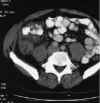 Figure
1. (click image to zoom) Inhomogeneously enhancing retrocecal mass with
extensive inflammation in right retroperitoneum and pericecal area. Figure
1. (click image to zoom) Inhomogeneously enhancing retrocecal mass with
extensive inflammation in right retroperitoneum and pericecal area.
Laparotomy
4 weeks after the diagnosis revealed an inflamed retrocecal appendix consistent
with appendicitis. The appendectomy was completed uneventfully, and the
patient's postoperative course was uncomplicated. Pathologic examination of the
appendix showed chronic inflammatory changes with diverticular formation
characteristic of diverticulitis (Fig 2).
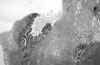 Figure
2. (click image to zoom) Figure
2. (click image to zoom)
Serosal
surface of appendix with fibrous thickening of wall and inflamed mucosa. Outer
muscular layer of appendix can be seen at lower right.
Discussion
Kelynack[6]
first described diverticulosis of the vermiform appendix in 1893. Since that
time, many others have proposed theories on the pathogenesis of the diverticula.
As with all intestinal diverticula, those found in the appendix can be
classified as congenital or acquired.
In
the congenital diverticulum, all layers of the bowel wall are present. This is
extremely rare, with fewer than 50 cases reported world-wide. Favara[7]
suggested a chromosomal basis for this lesion. In reviewing eight patients with
multiplecongenital appendiceal diverticula, seven were infants born with trisomy
D 13-15 syndrome. Other suggested mechanisms include appendiceal duplication,
local sacculations formed during appendiceal recanalization, epithelial
inclusion in the appendiceal wall, and traction. [8]The wall of the diverticulum
in the acquired cases contains only mucosa, submucosa, and serosa. Trollope and
Lindenauer[2] published a collective review in 1974 of the 1,373 known cases
along with a discussion of the most common theories of pathogenesis. The first
theory involves a post-appendicitis weakness of the bowel wall, leading to
ulceration and secondarily regenerated epithelium over the injured area.
Stout[9] reported the most likely noninflammatory theory in 1923.He
believed that appendiceal diverticula could be formed by a combination of
luminal obstruction and active muscular contraction. The obstruction, coupled
with the 1 to 2 mL of appendiceal secretions that are produced daily, is
augmented by contractions of the appendix in an effort to empty itself; the
result is a high enough pressure to cause diverticular formation or perforation.
[2] It is likely that in most cases multiple factors lead to the development of
acquired appendiceal
diverticulosis.
Progression
from diverticulosis to diverticulitis follows a partial or complete obstruction
of the lumen. This may be due to swelling of the mucosa, inflammation,
fecaliths, fibrous strictures, or torsion. [10-12] In the pre-antibiotic era,
most of these patients had a preoperative diagnosis of chronic appendicitis. The
pain is often described as insidious in nature, intermittent, and extended over
a long period.
Low-grade
fever and leukocytosis are commonly found. Anorexia, nausea, and vomiting are
usually absent. Most of the patients have had one or more admissions before the
operative admission.
Appendiceal
diverticulitis is an uncommon problem. It is also clear that the incidence is
greater than that generally appreciated. Since Trollope and Lindenauer's
original 1,373 cases 2 were reported, an additional 294
cases 13-24 have been discussed in the English language literature. The average
age of the appendiceal diverticulitis patient is 37 years compared with 19 years
in cases of appendicitis.[2] Both the congenital and the acquired types are more
common in men. Nearly all appendiceal diverticula are of the acquired type. Due
to the thinned wall, these diverticula are prone to perforate early in the presence of acute inflammation.[25]
Grossly, the proximal appendix usually appears normal but plunges into an
inflamed mass covered with fibrinous exudate. Nearly 60% of the diverticula are
located in the distal third of the appendix.[11] In a comparison of acute
diverticulitis to acute appendicitis, perforation was found to be more than four
times as likely in the diverticulitis group at 66%.[2] Another reported
complication of appendiceal diverticulosis is hemorrhage requiring
several units of blood transfusion.[26] In addition, multiple cases of
pseudomyxoma peritonei have been reported from appendiceal diverticula. [27,28]
The patients most likely to have appendiceal diverticulitis are those with
cystic fibrosis. In these patients, the diverticula arise at the site of a
penetrating artery.
Most
of the patients are adolescents, and their age averages 13 years. The total
incidence of appendiceal diverticulosis in cystic fibrosis patients from autopsy
data is 14%. For cystic fibrosis patients with abdominal surgery excluding
laparotomy for meconium ileus, this rises to a 43% incidence. [16] No current
diagnostic radiographic evaluations are available for appendiceal
diverticulosis. Due to the likelihood of complications, diverticulosis of the
appendix is a finding that radiologists stress.
[29] Computed tomography has become an increasingly
popular tool for cases of nonspecific right lower quadrant abdominal pain, and
in most cases it can be used to determine if the condition is a surgical or
nonsurgical problem. Computed tomography findings in cases of appendicitis can
include appendiceal swelling, pericecal inflammation, abscess, phlegmon, and
increased density in the pericecal fat.[30] In the case of our patient, the CT
image shows a large pericecal phlegmon without evidence of abscess formation. It
does not clearly identify the appendiceal diverticulum.
Treatment
of appendiceal diverticulitis can be appendectomy, cecectomy, or right
hemicolectomy, depending on intraoperative findings. If the induration extends
onto the cecum, it may be difficult to differentiate the inflammatory mass of
diverticulitis from that of a tumor. In one case, [19] a laparoscopic
appendectomy for appendiceal diverticulitis was safely completed without
difficulties.
Conclusion
Although
appendiceal diverticulitis is rare, clinicians should be aware of its
occurrence. It is likely that this disease is neglected due to its rarity and
because the exact pathogenesis is still incompletely understood. What makes
appendiceal diverticulosis an important clinical entity is that, when combined
with appendicitis, it can lead to early perforation. This is due to the lowered
ability of the thin wall of the acquired diverticulum to withstand the increased
intraluminal tension leading to a fourfold increase in perforation rates.
Preoperative CT cannot be used to diagnose appendiceal diverticulitis but will
often distinguish surgical from nonsurgical right lower quadrant inflammation.
Consideration should be given to laparoscopy for potential appendectomy rather
than laparotomy if appendiceal diverticulitis is suspected from the CT. During
intraoperative exploration, the appendix should be examined to rule out
appendiceal diverticula; if found, appendectomy should be done unless
contraindicated by other medical problems.
[ التالي ] [ الفهرس ] [ السابق ]
|