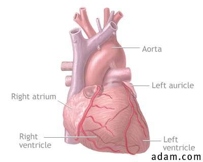
Abdominal aortic aneurysm

 
Aortic rupture, chest X-ray

 
Aortic dissection


An abnormal widening of the abdominal portion of the aorta (the major artery from the heart).

Alternative Names

aortic aneurysm (dissecting)

Definition

A condition in which there is bleeding into and along the wall of the aorta (the major artery from the heart); this condition may also involve abnormal widening or ballooning of the aorta (aneurysm).

Causes, Incidence and Risk Factors

Aortic dissection involves bleeding into and along the wall of the aorta (the major artery from the heart), most often because of a tear or damage to the inner wall of the artery. This usually occurs in the thoracic (chest) portion of the aorta but may occur in the abdominal portion also.
The exact cause is unknown, but risks include atherosclerosis and hypertension. Traumatic injury is a major cause of aortic dissection, especially blunt trauma to the chest as with the steering wheel of a car during an accident. Aortic dissection may also be associated with other injury, infection, congenital weakness of the aorta, collagen disorders such as Marfan's syndrome, pseudoxanthoma elastoma, Ehlers-Danlos syndrome, relapsing polychondritis, or abdominal aortic aneurysm. Pregnancy, valve disorders (including aortic insufficiency), and coarctation of the aorta may also be associated with aortic dissection.
Aortic dissection occurs in approximately 2 out of 10,000 people. It can affect anybody, but it is most often seen in men 40 to 70 years old.
 

Symptoms

Note: Symptoms may begin suddenly.
Additional symptoms that may be associated with this disease:

Signs And Tests

Listening with a stethoscope (auscultation) at the chest and abdomen may reveal a "blowing" murmur over the aorta, a heart murmur, or other abnormality. There may be decreased (weak) pulses in the upper extremities. There may be signs of tamponade or hypovolemia, or signs resembling acute MI. There may be signs of shock but with normal blood pressure.
Aortic dissection or aortic aneurysm may be revealed on:


Treatment

The goal of treatment is prevention of complications. Hospitalization is usually required.
Antihypertensives may be prescribed to reduce blood pressure. These may be given through a vein (intravenous). Analgesics may be needed for pain. Cardiac medications such as beta-blockers may reduce some of the symptoms.
Surgical repair or replacement of the section of aorta is curative.

Expectations (Prognosis)

Aortic dissection may be life threatening. The disorder is curable with surgical repair if it is performed before aortic rupture. Less than half of the patients with ruptured aorta survive.

Complications

  • bleeding from the aorta
  • aortic rupture causing rapid blood loss, shock, death
  • clot formation
  • insufficient circulation past the area of the dissection
  • irreversible kidney failure
  • stroke
  • myocardial infarction (tissue death)
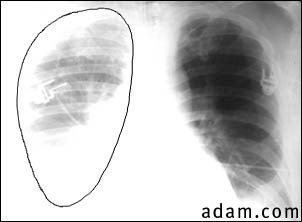
  •  Aortic rupture (a tear in the aorta, which is the major artery coming from the heart) can be seen on a chest X-ray. In this case, it was caused by a traumatic perforation of the thoracic aorta. This is how the X-ray appears when the chest is full of blood (right-sided hemothorax) seen here as cloudiness on the left side of the picture.
  • cardiac tamponade
  •  

Calling Your Health Care Provider

If symptoms indicate aortic dissection may be present or severe chest pain develops, call the local emergency number (such as 911) or go to the emergency room.
 

Prevention

Adequate treatment and control of atherosclerosis and hypertension may reduce risk. Use safety precautions to reduce the risk of injury. Many cases are not preventable.


Infective endocarditis

 
Aortic rupture, chest X-ray

 
Thoracic aortic aneurysm


Alternative Names

aneurysm - thoracic aortic; aortic aneurysm (thoracic); syphilitic aneurysm

Definition

A localized dilation of the wall of the thoracic aorta caused by atherosclerosis, hypertension or, less commonly, syphilis. Syphilitic aneurysms nearly always occur at the thoracic aorta, whereas more common aneurysms, due to atherosclerosis, nearly always occur in the abdominal aorta. This condition is rare in children.


Aortic arch syndrome

Alternative Names

carotid artery occlusion syndrome; subclavian artery occlusive syndrome; subclavian steal syndrome; vertebral-basilar artery occlusive syndrome

Definition

A pattern of signs and symptoms associated with abnormalities in the structure of the arteries that extend from the aortic arch (the top portion of the aorta). Symptoms include various neurologic changes, blood pressure changes, reduced pulse, and others. This syndrome is most often associated with atherosclerosis, trauma, blood clots, or congenital abnormalities of vessel structure.


Aortic insufficiency

Alternative Names

aortic regurgitation; aortic valve prolapse

Definition

A condition in which there is weakening or ballooning of the aortic heart valve, causing the valve to fail to close tightly with subsequent back flow of blood into the ventricle.

Causes, Incidence and Risk Factors

Aortic insufficiency is a heart valve disease where the aortic valve no longer functions adequately to control the flow of blood from the left ventricle into the aorta.
Aortic insufficiency can result from any condition that weakens the aortic valve. Rheumatic fever used to be the primary cause of aortic insufficiency. With antibiotic treatment of rheumatic fever, other causes are being seen with increasing frequency. These include congenital conditions, endocarditis, hypertension, Marfan's syndrome, aortic dissection, ankylosing spondylitis, Reiter's syndrome, syphilis, and other disorders.
The disorder affects approximately 5 out of 10,000 people. It is most common in men 30 to 60 years old.
 

Symptoms

Note: Commonly, aortic insufficiency shows no symptoms for many years. Symptoms may then occur gradually or suddenly.

Signs And Tests

Auscultation (listening to the chest with a stethoscope) shows a heart murmur. Palpation (examination by hand) may show hyperdynamic (very forceful) beating of the heart. Pulse pressure (the difference between systolic and diastolic blood pressure) may be widened to more than 60, and diastolic blood pressure may be low. There may be signs of pulmonary edema.
Aortic insufficiency may be seen on:

An ECG test or Chest X-ray may show left ventricle enlargement.
Lab tests are nondiagnostic for aortic insufficiency, but may they be used to rule out other disorders or causative factors.
 

Treatment

If no symptoms, or if symptoms are mild, the disorder may only require observation. If symptoms are severe, hospitalization may be required. Medications such as diuretics or digoxin may be used to stabilize the condition. These medications may also be used in people with mild symptoms to prevent the symptoms from worsening. Moderate activity restriction may be recommended.
Surgical repair or replacement of the aortic valve corrects aortic insufficiency. Surgical repair of the aorta may be required if insufficiency is caused by disorders of the aorta.

Expectations (Prognosis)

Aortic insufficiency is curable with surgical repair. The person may become symptom-free unless complications develop.

Complications

Calling Your Health Care Provider

Call for an appointment with your health care provider if symptoms indicate aortic insufficiency may be present.
Call your health care provider if symptoms worsen or new symptoms develop in a person with aortic insufficiency; especially chest pain, difficulty breathing, or edema (swelling).
 

Prevention

Aortic insufficiency cannot be prevented but some of the complications can be.
Notify the health care provider or dentist about any history of heart valve disease before treatment for any condition. Any dental work, including cleaning, and any invasive procedure, can introduce bacteria into the bloodstream. This bacteria can infect a weakened valve causing endocarditis.
Follow the provider's treatment recommended for conditions that may cause valve disease. Treat strep infections promptly to prevent rheumatic fever. Notify the provider if there is a family history of congenital heart diseases.
 

This is an illustration of the exterior of the heart. The arteries are colored blue and represent blood that is flowing into the heart. The veins are red and indicate blood flow out from the heart. The chambers are also labeled.


This is a cross-sectional view of the interior of the heart. The valves, chambers, and associated vessels are labeled.


Aortic stenosis

Alternative Names

aortic valve stenosis

Definition

A narrowing or obstruction of the aortic heart valve, causing it to not open properly and to obstruct the flow of blood from the left ventricle to the aorta.

Causes, Incidence and Risk Factors

Aortic stenosis is caused by many disorders. One cause is rheumatic fever. Other causes include calcification of the valve and congenital abnormalities. There may be a history of other valve diseases, coronary artery disease, or heart murmur.
Aortic stenosis occurs in approximately 5 out of 10,000 people. It is more common among men. Symptoms often do not appear until middle age or older.
 

Symptoms

Note: Aortic stenosis may show no symptoms until late in the course of the disease.
Additional symptoms that may be associated with this disease:

Signs And Tests

Examination shows a palpable chest thrill or heave (vibration or movement felt by holding the hand over the heart). There is almost always a heart murmur, click, or other abnormal sounds on auscultation (examination of the chest with a stethoscope). There may be faint pulses or changes in the quality of the pulse in the neck (the change in pulse is known as pulsus tardus). Blood pressure may be low.
Aortic stenosis and/or enlargement of the left ventricle may be revealed on:

An ECG may show left-ventricle enlargement or arrhythmias (unusual pattern of heart beats) such as ventricular tachycardia or sinus bradycardia.
This disease may also alter the results of the following:


Treatment

If there are no symptoms or symptoms are mild, only observation may be required. If symptoms are mild to severe, hospitalization may be required. Medications may include diuretics, digoxin, and other medications to control heart failure. Symptomatic people may be advised to avoid strenuous physical activity. People with symptoms of aortic stenosis (difficulty breathing, chest pain, syncope) should have a physical exam every 6 to 12 months, and an ECG performed every 1 to 3 years.
Surgical repair or replacement of the valve is the preferred treatment for symptomatic aortic stenosis.

Expectations (Prognosis)

Aortic stenosis is curable with surgical repair, although there may be a continued risk for arrhythmias. The person may be symptom-free until complications develop. Without surgery, probable outcome is poor if there are signs of angina or heart failure.

Complications

Calling Your Health Care Provider

Call for an appointment with your health care provider if symptoms indicate aortic stenosis may be present.
Call your health care provider if aortic stenosis has been diagnosed and symptoms worsen or new symptoms develop.
 

Prevention

Aortic stenosis cannot be prevented, but some of the complications can be.
Notify the health care provider or dentist about any history of heart valve disease before treatment for any condition. Any dental work, including cleaning, and any invasive procedure, can introduce bacteria into the bloodstream. This bacteria can infect a weakened valve causing endocarditis.
Follow the provider's treatment recommended for conditions that may cause valve disease. Treat strep infections promptly to prevent rheumatic fever. Notify the provider if there is a family history of congenital heart diseases.


Marfan's syndrome


Pectus excavatum is a condition in which the "breast bone" (sternum) appears sunken and the chest concave. It is sometimes called "funnel chest". The majority of these cases are not associated with any other condition (isolated findings). However, some syndromes include pectus excavatum.

Definition

An inheritable disorder that affects the skeletal system, cardiovascular system, eyes, and to some extent the central nervous system.

Causes, Incidence and Risk Factors

Marfan's syndrome is inherited as an autosomal dominant trait. Marfan's syndrome may appear in previously unaffected families as a spontaneous new mutation. The incidence is approximately 1 out of 10,000 people.
Marfan's may cause many skeletal defects but is most notably recognized for a tall, lanky frame with long arms and spider-fingers (arachnodactyly), and chest abnormalities (pectus excavatum or pectus carinatum).
Common eye problems are nearsightedness (myopia) and dislocation of the lens of the eye. The white of the eye (sclera) may appear bluish in Marfan's syndrome as it does in several other diseases with skeletal abnormalities.
The most significant of the defects in the syndrome are cardiovascular abnormalities, which may include enlargement (dilatation) of the base of the aorta (aortic root), with aortic regurgitation and prolapse of the mitral valve. Affected people may develop a dissecting aortic aneurysm.
 

Symptoms

  • family history of Marfan's syndrome
  • long, lanky frame
  • long, thin limbs
  • armspan significantly greater than body height
  • long, spidery fingers (arachnodactyly)
  • funnel chest (pectus excavatum) or pigeon breast (pectus carinatum)
  • scoliosis
  • visual difficulties
  • flat feet
  • learning disability/school problems
  • thin narrow face
  • micrognathia (small lower jaw)
  • coloboma of iris
  • hypotonia

Signs And Tests

Physical examination may show:

There may also be signs of:


Treatment

There is not a single curative treatment for this condition.
Visual defects should be corrected where possible. Care should be taken to prevent the development of scoliosis, especially during adolescence.
The cardiovascular abnormalities are the most worrisome. People with Marfan's should have preventive antibiotics before dental procedures in order to prevent endocarditis. Pregnancy in individuals with Marfan's needs to be monitored very closely because of the increased stress on the heart and aorta. Children should not be involved in maximal exercise programs or sports because of concern for aortic aneurysm.

Expectations (Prognosis)

Lifespan is shortened somewhat because of cardiovascular complications. The survival is into the 40s but may be extended with good care and heart surgery.

Complications

Calling Your Health Care Provider

You have a family history of Marfan's syndrome and are considering having children. Also call if you child has symptoms suggestive of this syndrome.
 

Prevention

Spontaneous new mutations leading to Marfan's (less than 1/3 of cases) cannot be prevented. However, for those prospective parents with a family history of Marfan's syndrome, genetic counseling is recommended.