 545x700
24-bit color JPEG, 69069 bytes
545x700
24-bit color JPEG, 69069 bytesLIST
OF IMAGES OF DIFFERENT MODALITIES WITH THEIR DATA PROPERTIES
COLOR
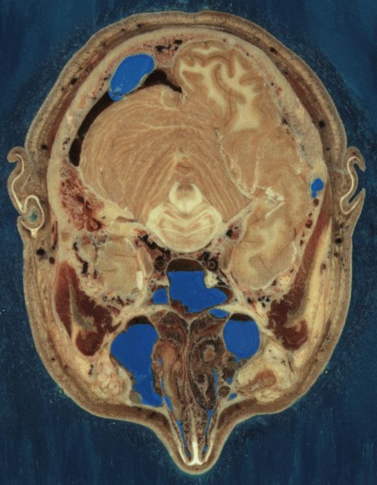
CRYOSECTIONS
545x700
24-bit color JPEG, 69069 bytes
Section through Visible Human Male - head, including cerebellum, cerebral cortex, brainstem, nasal passages (from Head subset)
 786x459
24-bit color JPEG, 69920 bytes
786x459
24-bit color JPEG, 69920 bytes
Section through Visible Human Male - abdomen, including large and small intestines, spinal column, musculature, subcutaneous fat (from Abdomen subset)
 749x450
24-bit color JPEG, 64159 bytes
749x450
24-bit color JPEG, 64159 bytes
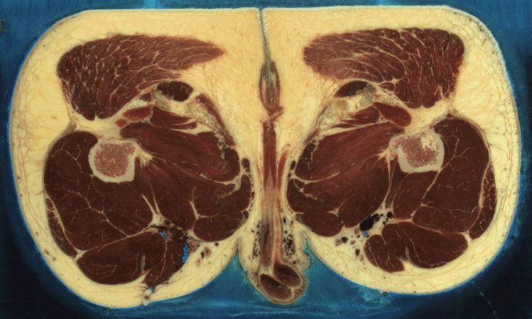
Section through Visible Human Male - upper thigh below femoral head, including prominent musculature, part of male reproductive system (from Pelvis subset)
 730x399
24-bit color JPEG, 51365 bytes
730x399
24-bit color JPEG, 51365 bytes
Section through Visible Human Male - knee, including
patella (from Thigh subset)
CASE
STUDY
1)
Fracture of the temporal bone.


Following trauma the 31-year-old
patient was admitted with conductive hearing disorder on the right side.
Otomicroscopy showed a fracture of the temporal bone.
Computed Tomography was performed
using a Multi-Slice-CT (GE CT Light Speed QX/i). CT was obtained with 1.25 mm
collimation, exposure duration of 7.8 seconds and pitch of 3 (140 kV, 40 mA).
Reconstructed images were generated using a bone reconstruction algorithm
at 0.3-mm interval and 9.6 cm field of view. CT revealed a fracture of the
temporal bone and dislocation of the incudomallear and incudostapedial joint.
The dislocation of the incus was caused by impingement of a fracture fragment.
Source: Department of
Otorhinolaryngology, Hannover Medical School
2) Conjoined
Twins with Complex anatomy.

A 14-week Ultrasound revealed a set of twins joined at the chest and abdomen. The mother was 29-year-old woman from Puerto Rico. The twins shared a single heart that was physically located in the chest of one of the fetuses. This heart circulated blood between both fetuses. A smaller non-functional heart was present in the second twin. The blood flow was complicated because the placenta was required to be present in order to complete the circulation path. Without the placenta, blood flow would be inadequate to keep either twin alive.
MRI
IMAGE.
Coronal wrist, 7 cm FOV, 1.5 mm slice thickness, SE, 256x256 matrix,
TR/TE (Repetition time/Echo time)= 2000/27 ms, 1 Number of Excitation.
|
|
|
||
|
|
DICOM Format Implicit Little MR MONOCHROME2 Resolution 256x256 pixels [16, 16 | 15], PR=1 G.E. Med. System Source: S.Barre’ Medical Imaging Samples |
|
|
CT
SCANS PRIOR TO FREEZING

Specimen from the Visible Human Male - Head subset
Resolution: 277x338 Pixels, 10740 bytes
Bit depth: 8-bit grayscale
Format: JPEG
Specimen from the Visible Human Male - Head subset
Source: Projects based on the Visible Human Data Set,
National Library of Medicine (USA)

Specimen from the Visible Human Male - Abdomen subset
Resolution: 511x354 pixels, 15722 bytes
Bit depth: 8-bit grayscale
Format: JPEG
Specimen from the Visible Human Male - Abdomen subset
Source: Projects based on the Visible Human Data Set, National Library of Medicine (USA)
CT
SCANS AFTER FREEZING

Specimen from the Visible Human Male - Head subset
Resolution: 353x451 Pixels, 16811 bytes
Bit depth: 8-bit grayscale
Format: JPEG
Source: Projects based on the Visible Human Data Set, National Library of Medicine (USA)

Specimen from the Visible Human Male - Abdomen subset
Resolution: 512x340 Pixels, 16548 bytes
Bit depth: 8-bit grayscale
Format: JPEG
Source: Projects based on the Visible Human Data Set, National Library of Medicine (USA)
Spiral
CT scan
(2-mm collimation, pitch of 2, 24% iodinated contrast agent) obtained at the level of the lower lobes in a 70-year-old patient with a previous history of severe chronic obstructive pulmonary disease and acute dyspnea at the time of diagnosis. There is a partial filling defect (arrow) at the level of a subsegmental branch of the anterior segmental artery of the right lower lobe.
Source: Department of Radiology, Hospital Calmette,
Boulevard Jules Leclerc.

Patient referred for abdominal pain.
This is an axial slice of mid abdomen, across the kidneys.
Patient referred for abdominal pain.
Slice Thickness: 3mm
Resolution: 512x 512 pixels, 25k bytes
Bit depth: 12–bit grayscale
Format: JPEG
Source: Changi General Hospital Radiologist.

This is an image of the sigmoid colon after administration of barium contrast and air.
Patient referred for colon cancer.
Resolution: 512 x 512 Pixels, 26k bytes
Bit depth: 10-bit grayscale
Format: JPEG
Source: Changi General Hospital Radiologist.
This is a scan of the uterus, transverse section.
Patient referred for ovarian cysts.
Resolution: 512x 512 Pixels, 7K bytes
Bit depth: 10-bit grayscale
Format: JPEG
Source: Changi General Hospital Radiologist.
