Ocular Surgery News
a SLACK Incorporated newspaper

Refractive Surgery

Improvement in visual quality may be obtained with Intacs implantation associated with LASIK

By re-establishing physiological corneal asphericity, Intacs corneal ring segments improve visual acuity and contrast sensitivity, reducing glare and halo effects.

by Michela Cimberle


December 15, 1999

MILAN, Italy — Intacs intrastromal corneal ring segments (KeraVision, Fremont, Calif.) implantation associated with laser in situ keratomileusis (LASIK) considerably reduces undercorrection, prevents regression and allows myopic correction over 15 D, according to Carlo Lovisolo, MD, of the Vista Vision Eye Center, Milan. The surgical plan is: down-up LASIK, 240 µm safety residual bed; after 3 months, Intacs implantation through superior incision placed in the hinge area.

“The technique is safe, precise and easily adjustable. It is easy to learn and has a very good predictability,” he said.

His experience of more than 10 months of follow-up shows that Intacs implantation does not interfere with the clinical and topographic stability of the LASIK flap.

The flattening effect of Intacs on the anterior corneal surface is calculated by the nomograms that are normally used for low myopic correction, and the refractive result is easily adjustable.

“All you need to do is to change the implantation with segments of different thickness, and you can remove them whenever needed,” Dr. Lovisolo said. This offers patients around presbyopic age the option of monovision, taking the nondominant eye back to slight myopia.

[bar]
An unexpected bonus

“To our surprise, we observed that quality of vision improves tremendously; all patients previously treated with LASIK, photorefractive keratectomy (PRK) or radial keratotomy (RK), and later implanted with Intacs, gained one, or even two, best corrected visual acuity lines, and decisively improved their mesopic contrast sensitivity,” Dr. Lovisolo said.

The reasons for this phenomenon are not entirely clear. According to the surgeon, they could be related to three considerations:

  • Intacs implantation does not induce astigmatism. On the contrary, a correct positioning of the incision and suture can, in many cases, correct the astigmatism.
  • The functional optical zone is considerably enlarged.
  • The physiologic aspheric shape of the cornea, with a more pronounced curvature in the center than in the periphery, is often re-established.

The anterior corneal surface is the most important element in determining the power of the optical system of the eye (more than 80%). As such, it also is the main source of optical aberrations, and has a significant influence on the functional performance of our visual system.

A number of studies have confirmed that prolate asphericity (more pronounced curvature in the center than in the periphery) is the ideal shape of the anterior corneal surface, in comparison with other types of asphericity (paraboloidal, conical, hyperbolical, oblate ellipsoidal).

“The asphericity of a section is determined by mathematical calculation of the curvature variations between the apex and the periphery,” Dr. Lovisolo explained. “If the surface flattens from the apex to the periphery, the curve is said to be prolate and the ‘shape factor’ [p] is positive. Vice versa, the curve is said to be oblate, and the shape factor negative, when the surface curves from the apex to the periphery. Also corneal topography confirms that the physiological curvature of the cornea is elliptic and prolate. This conformation reduces spherical aberration and determines the quality of visual performance.”

Surgical correction of myopia (with LASIK, PRK or RK) alters, and often inverts, corneal asphericity. Authoritative surgeons believe that such an anti-physiological effect may be the cause of patients’ complaints such as glare, starburst and reduced contrast sensitivity in mesopic conditions.

“Intacs implantation, on the contrary, increases the positive asphericity of the anterior corneal surface,” Dr. Lovisolo said. “When dealing with eyes that have been previously treated with LASIK, PRK or RK, the implantation is able to invert the quotient of asphericity [Q], re-establishing more physiological visual conditions.”

[bar]
Measuring spherical variations

In order to measure the spherical variations of the cornea before treatment, after photoablation and after Intacs implantation, Dr. Lovisolo uses three indices of asphericity: eccentricity (e), quotient (Q) and shape factor (p), which are related as follows:

“The Q value is negative in normal corneas (ranging between –0.01 and –0.61. Average –0.26),” Dr. Lovisolo explained. “The average postoperative values after LASIK or PRK depend on ablation quantity, amplitude of optical zones and progression of transition zones. They are generally major than 1, and in EyeSys technology (Holladay Diagnostic Summary), they stop at 1.99 de fault value. After Intacs implantation, Q is back to slightly positive values (between 0 and 0.3).”

photographphotograph
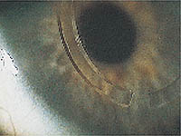
Different enlargements of Intacs in situ. The eye had previously undergone myopic LASIK. Observe the margin of primary keratectomy.
topography
Pre-LASIK corneal topography (Holladay Diagnostic Summary, EyeSys) of right eye in 50-year-old patient.
topography
Same eye, corneal topography 6 months after LASIK.
topography
Same eye, corneal topography 4 months after Intacs implantation. Observe the values of Q (from –0.24 to +1.99 to +0.01), of SRI (from 100% to 70% to 80%) and potential corneal visual acuity (from 20/10 to 20/32 to 20/25).
videokeratoscopy
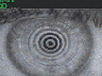
Two months postop videokeratoscopy.
tangential map
Tangential map and difference map of –15 sphere –1 cylinder corrections achieved with LASIK and Intacs. Note the amplitude of the optical zone induced by Intacs ring segments.
tangential map
Tangential map and difference map of –15 sphere –1 cylinder corrections achieved with LASIK and Intacs. Note the amplitude of the optical zone induced by Intacs ring segments.
tangential map
Tangential map and difference map of –15 sphere –1 cylinder corrections achieved with LASIK and Intacs. Note the amplitude of the optical zone induced by Intacs ring segments.
tangential
Orbscan corneal tomography. Preoperative elevation map of the anterior corneal surface.
elevation map
Six months after LASIK orbscan elevation map.
tomography
Two months Orbscan tomography after Intacs implantation.
For Your Information:
  • Carlo Lovisolo, MD, can be reached at Via Formentini 1, 20121 Milan, Italy; (39) 02-805-7388; fax: (39) 02-864-52896; e-mail: loviseye@tin.it. He also can be reached at Vista Vision Eye Center, Via Rizzo 8, Milan, Italy; (39) 02-38005605; fax: (39) 02-38008037. Dr. Lovisolo has no direct financial interest in any of the products mentioned in this article, nor is he a paid consultant for any companies mentioned.
  • For more information on the Intacs intrastromal corneal ring segments, contact KeraVision Inc., 48630 Milmont Drive, Fremont, CA 94538-7353; (510) 353-3000; fax: (510) 353-3030.

Copyright 1999, SLACK Incorporated. Revised 9 December 1999.
Source Site