
a SLACK Incorporated newspaper
Editor’s note: This is the first in a series of articles on experiences with the Kera Vision (Fremont, Calif.) Intacs. In this article, Daniel S. Durrie, MD, discusses his surgical maneuver for implanting the Intacs.
by Daniel S. Durrie, MDAugust 15, 1999 Laser in situ keratomileusis (LASIK), though highly effective, really was designed for moderate to high myopia and to correct astigmatism. If one goes back just 2 years ago, few surgeons were routinely performing LASIK for patients with 1 D and 2 D of myopia. Only recently have more surgeons become comfortable enough with LASIK that they have worked down into the low levels of myopia. However, now there is an alternative to LASIK for patients with low myopia. KeraVision Inc.’s (Fremont, Calif.) Intacs intrastromal corneal ring segments are specifically designed for low-level myopia. Patients like the idea that this procedure has been designed and tested specifically for them. Surgeons like the procedure because it is easily reversible and takes no more time than LASIK.
|
|||
  |
|||
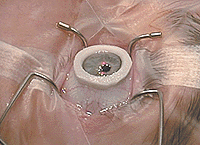
| There is a clockwise and counterclockwise ring. They are placed in the channel with a twisting or dialing motion. | |||
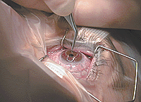
| The second step of the implantation procedure entails creating the channels in the stroma in a clockwise and counterclockwise direction. This is done by placing a special suction ring, made by KeraVision, on the cornea and centering it (Figure 6). The suction ring has a centering de vice and is well designed. It does not slip or slide and maintains a good grip on the eye. After suction is released, we begin the third stage of the procedure, which is the easiest. In this step, the Intacs segments are placed into the channel (Figures 7a, 7b and 7c). There is a clockwise and counterclockwise ring. Each ring is designated as such in the packaging so that you know in which direction to go. Those are placed in the channel with a twisting or dialing motion.
|
|||
  |
|||
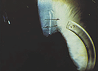
| Dr. Durrie advises physicians with minimal Intacs experience to suture the incision with a single, 11-0 nylon suture. | |||
| There is still a debate over whether the incision should be closed with a suture — as we did in the clinical trials — or left open. Physicians with minimal Intacs experience should suture the incision with a single, 11-0 nylon suture (Figures 8a and 8b). This is to approximate the anterior leaflets of the incision. Suturing should be continued until you are completely comfortable with the procedure. Currently, I am involved in a randomized, prospective study comparing sutured with non-sutured Intacs eyes. In Canada and Europe, when the physicians stopped using sutures, outcomes suffered; however, we are not sure why. I suspect that the incision communicates with the channel during healing. If the wound is even open slightly during the first 1 to 2 weeks postop, tears can enter the channel and could cause variability in wound healing or some variation in vision throughout the day. One of the drawbacks to the suture is that if you tie it too tightly, you will induce astigmatism. Sutures should be re moved approximately 1 month after surgery.
|
|||
 ---Dr. Durrie uses a slit, 3M 1020 drape and
a wire lid speculum — the same format he uses for LASIK.
---Dr. Durrie uses a slit, 3M 1020 drape and
a wire lid speculum — the same format he uses for LASIK.
 ---The KeraVision marker is placed around the
geometric center of the cornea.
---The KeraVision marker is placed around the
geometric center of the cornea.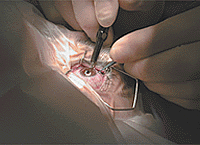 ---The incision is made with a
diamond blade to two-thirds of the depth of the corneal thickness at the
location of the incision, which needs to be measured intraoperatively.
---The incision is made with a
diamond blade to two-thirds of the depth of the corneal thickness at the
location of the incision, which needs to be measured intraoperatively.
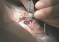 ---Dr. Durrie grasps the edge of the wound
with the forceps and looks through the bottom of the incision when starting the
pocket with the Sinskey hook.
---Dr. Durrie grasps the edge of the wound
with the forceps and looks through the bottom of the incision when starting the
pocket with the Sinskey hook.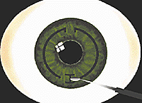 ---The pocket is extended with a stromal
spreader, which allows room for a 1.8 mm wide channel, 2 mm in length and
extending clockwise and counterclockwise from the original incision.
---The pocket is extended with a stromal
spreader, which allows room for a 1.8 mm wide channel, 2 mm in length and
extending clockwise and counterclockwise from the original incision.
 ---Place a special suction ring, made by
KeraVision, on the cornea and center it.
---Place a special suction ring, made by
KeraVision, on the cornea and center it.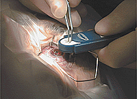 ---After suction is released, the Intacs
segments are placed into the channel.
---After suction is released, the Intacs
segments are placed into the channel.