
a SLACK Incorporated newspaper
April 1998
 ---Preop
corneal topography of a keratoconus patient selected to receive ICRS
implant.
---Preop
corneal topography of a keratoconus patient selected to receive ICRS
implant.
BREST, France - Because of their safety, reversibility and predictability, intrastromal corneal ring segments (ICRS) are gaining wide acceptance as viable alternatives to refractive laser surgery for patients with myopia. Now, research by Joseph Colin, MD, a professor of ophthalmology here at the University of Brest, suggests that ICRS may also benefit patients with keratoconus.
When keratoconus thins corneal tissue, the structure is often left weak and unstable. Pressure from within the eye causes the cornea to bulge outward, causing severe myopia in some patients. Penetrating keratoplasty (PK) is commonly used to restore structural integrity and visual acuity in patients with advanced keratoconus, but transplantation incurs additional risk. Dr. Colin's work suggests that the ICRS may be used to flatten and strengthen a clear, but otherwise failing, cornea.
"ICRS [implantation] could be an interesting surgical alternative to delay or avoid PK in patients with clear corneal keratoconus and contact lens intolerance," he said.
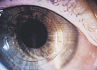
 ---Slit-lamp
examination of ICRS placed in keratoconic eye.
---Slit-lamp
examination of ICRS placed in keratoconic eye.
Dr. Colin, along with David J. Schanzlin, MD, a professor of ophthalmology at the University of California at San Diego, and Beatrice Cochener, MD, in private practice in Brest, France, sought to evaluate the technique on a 22-year-old woman with a 7-year history of bilateral keratoconus. Three years from the time of diagnosis, the patient wore spectacles with an acceptable level of best-corrected visual acuity, but after that period, she was fitted with contact lenses. Her right eye, which underwent the procedure, retained a clear cornea, but had become contact lens intolerant 9 months before being referred to Dr. Colin for PK. At the time of surgery, the patient had right-eye, central corneal thickness of 515 µm.
Dr. Colin said the ICRS procedure for keratoconus varies little from the procedure for myopia. After the patient was administered topical anesthesia, a diamond blade was used to create a 1.8-mm incision, perpendicular to the periphery of the cornea, to a depth of approximately 68% of the peripheral corneal thickness, which, in this case, varied from 600 µm to 630 µm. The incision was then laterally separated at its base to prepare an entry incision on each side.
A stromal pocket was created with a Suarez spreader, and then two semi-channels, in the inferior and superior positions, were created for the rings. Ring segments were "dialed" into position, and the incision was closed with 10-0 nylon sutures that were left loose so as to not induce astigmatism. Dr. Schanzlin said procedures typically take about 15 minutes.
In this particular case, Dr. Colin said that a 0.45-mm segment, the thickest available, was selected to flatten and strengthen the inferior inner stroma, while a 0.35-mm superior segment was used to decrease postoperative astigmatism.
Postoperative management was also similar to that when the ICRS is used for the correction of myopia. A topical antibiotic and corticosteroids were administered several times daily. Sutures were removed 4 weeks postop.
Seven days postop, refraction was 20° -3.75 D +0.5 D, and uncorrected visual acuity was 0.7. Four months later, visual acuity and refraction were unchanged. Corneal topography revealed a decrease in inferior corneal steepening.
 ---Postop
topography of same keratoconus patient with ICRS in place.
---Postop
topography of same keratoconus patient with ICRS in place.
"Our first case suggests that the ICRS may be efficient to decrease corneal steepening induced by keratoconus and to decrease corneal astigmatism; however, some questions remain," Dr. Colin said. Researchers are uncertain whether soft, keratoconic stromal tissue reacts to ICRS implantation the way normal myopic corneas do. There are also unanswered questions about the best way to select the thickness of superior and inferior segments, to decrease residual or induced asymmetrical corneal astigmatism.
Dr. Colin said the soft corneal tissue of keratoconus patients poses potential problems during implantation.
"In addition to testing the reaction of soft tissue to the rings, we need to evaluate the risk of perforation," he said.
Dr. Colin presented his case study at the International Society of Refractive Surgery meeting in San Francisco.
For Your Information:
- Joseph Colin, MD, can be contacted at Ophthalmologie, Chr Morvan Hospital, Brest 29299, France; ++(98) 22-34-40; fax: ++(98) 46-49-70; e-mail: Joseph.Colin@univ.brest.fr. Dr. Colin has no direct financial interest in the products mentioned in this article, nor is he a paid consultant for any companies mentioned.