

HISTOLOGY AND ANATOMICAL IMAGES
| Pituitary Gland [sagittal section] | Pituitary Gland [sagittal section, labeled] | Cross Section of Human Brain |
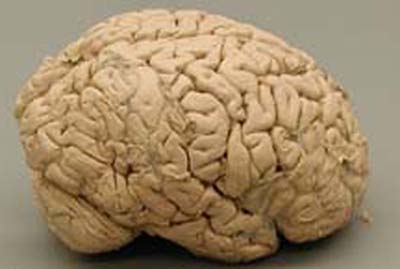
| Chest X-Ray | Lungs | Human Brain (lateral view) |
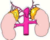
| Adrenal Gland Diagrammed | Pituitary Gland (labeled diagram) | Pituitary Gland |
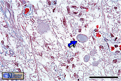
| Simple Squamous Epithelium | Trachea illustrating an example of pseudostratified columnar epithelium | Posterior Lobe of Pituitary Showing Herring bodies (blue arrow) |
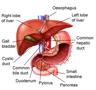
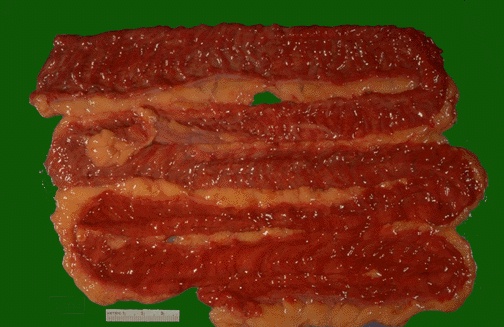
| Human liver (labeled) | Polycystic Kidneys | Intestinal Evisceration |
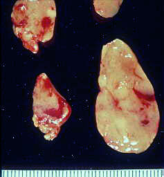
| Coronary artery thrombus (100% blockage) | Pulmonary edema (from autopsy) | Parathyroid hyperplasia |
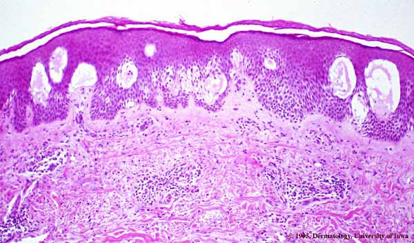
| Metastatic lung carcinoma | Acute contact dermatitis | Hyperplastic Prostate |
| Normal Spleen (cross section) | Thyroid Gland (Grave's Disease) | Normal Thyroid (histology) |
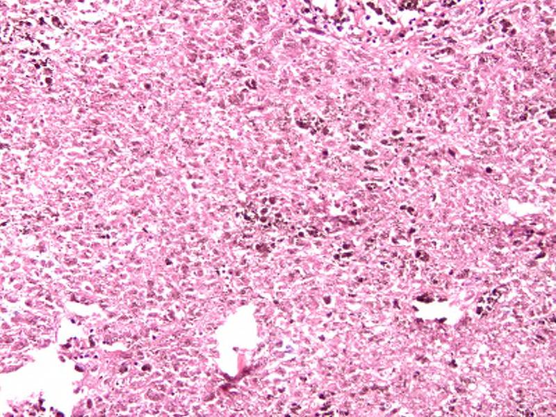
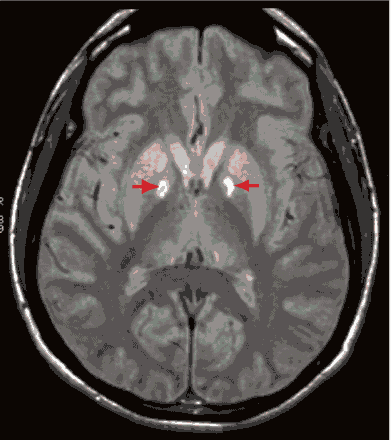
| Ischemic enteritis | Gangrenous Colon | Infant with lymphangioma | Liver histology, showing hemochromatosis | Neurofibrillary tangles, as seen in Alzheimer's Disease | Globus pallidus lesions |
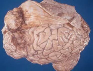
| Adenomatous colon polyp | Meningioma | Cerebral Infarction |
This page last updated on
August 15, 2009