| The Chest | ||
| Chest X-rays
|
As I mentioned on the
diagnostic One thing that does confuse a lot of patients about these is the PA, or frontal view, since the patient is facing away from the x-ray tube when this one is taken. Truthfully, referring to this as a frontal view is not correct, and most technologists only use this term because it is a term that they feel their patients will understand. The reason the patient is facing the chest board instead of the x-ray tube for this view is to place the heart closer to the x-ray film, thus reducing the magnification of the heart to a point where fairly accurate measurements of the heart can be obtained. These measurements are used to check for various heart conditions, such as CHF (congestive heart failure) and Cardiomegaly (basically an enlarged heart). A chest x-ray will also demonstrate a number of lung problems including such problems as: Bronchitis, Pneumonia, Asthma, Pnemothorax (air trapped between the lungs and the chest wall), and fluid in the lungs as a result of CHF. The lateral, or side view of the chest is obtained to see whether a problem is located nearer to the front of the chest or the back, and to see if there is anything behind the heart that would not show up on the PA view. The Technologists doing your x-rays will normally take the films after telling you to take in a deep breath and hold it. This expands the lungs to their fullest and pushes the diaphragm down, making it easier for the Radiologist who will read your x-rays to see everything. Not very long ago, when your doctor suspected a Pneumothorax of some kind the technologist would do two PA’s. One would be done in the normal manner with you taking in a deep breath and holding it, and the other would be done with you blowing your breath all the way out and not breathing in again until after the x-ray had been taken. The reason for the second film, called an exporation or expiratory film, was to more clearly demonstrate the pneumothorax which would increase in size temporarily as you blew your breath out. However today many doctors feel that a pneumothorax small enough to be missed on a normal chest film will more than likely repair itself if the patient just takes it easy for a while. Thus, unless expressly ordered by your doctor, most facilities only do a routine, deep breath in and hold type of chest x-ray. Another structure located in the chest region is the rib cage. For x-rays of the ribs, the patient preporation is limited to the same clothing considerations as a routine chest exam. The exam itself consists of taking three or four films, two frontal shots and one or two obliques, or angled pictures, depending on where the patient is hurting. The two frontal pictures are taken because the ribs cover not only your heart and lungs, but parts of your liver, stomach, and kidneys as well. The reason two shots are required on most people is that a setting or technique that will demonstrate the upper ribs that enclose your lungs will be too light to adequately show the ribs overlaying the upper abdomen (liver, stomach, and kideys), while a technique that will show the lower ribs will most likely be too dark to see any details of the upper ribs. The reason the oblique views are obtained is that since the ribs are curved, a straight side shot would not show any injuries or fractures on the curved sections well enough. Beyond this however, there tends to be a wide latitude on just how rib series (another name for the study) are done. Some facilities will x-ray only the affected side, others will follow the suggestions put forth in Merrill's Atlas of Radiologic Procedures more carefully and do both the side that hurts and the side that doesn't. Some facilities will just do films aimed at showing the details of the ribs, others will also include a standard single view of the chest. And some have done away with doing rib series completely. Since most rib fractures cannot be splinted in any viable way that will not also put the patient at risk of developing mild pneumonia as a result of constricting the amount of air that can be moved in and out of the lungs, these facilities will just do a chest x-ray to be sure that any fractured ribs have not caused either a pneumothorax or a hemothorax. The last part of the chest which may be x-rayed is the sternum, or breast bone. The sternum is perhaps one of the hardest bones in the body to obtain a good, high quality film of. There are two main reasons for this. First, since the sternum is directly in front of the spine, one cannot get a frontal view of it by shoting strait down at it. Instead the patient must be rolled slightly to allow the image of the sternum to show up beside the spine. The second problem is that the sternum, while very tough, is also very thin. As a result, if the settings used by the technologist is even slightly off, you won't be able to see the sternum at all. Yet the same settings that will allow you to see the sternum beautifully will also show up all the ribs that connect to the sternum. The way we deal with this is to use what we call a "breathing technique", which basically means that we have the patient continue to breath in a normal way while using settings on the x-ray machine that cause the x-ray beam to spread it self out over a relatively long period of time, say 30 seconds to two minutes instead of the more usual tenth of a second or so. This causes the ribs to move in such a way as to be blurred on the x-ray, allowing a better look at the sternum. |
|
[Home] [Cat Scan] [MRI] [Mammography] [Sign Guestbook] [Bio] |
||
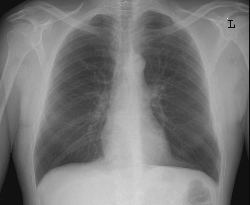 x-ray
contents page, the most commonly performed x-ray is a Chest x-ray. This exam consists of a
PA, or frontal view, or a PA and a Lateral, or side view. Since these films will be done
primarily to see the lungs and heart shadows (neither really shows up on x-rays, but parts
will cast shadow that do) the patient must not have anything on from the waist up that
will show up on x-rays. For example, if you are the patient, you must not have on:
necklasses, certain types of bras (with some bras I can even tell you the brand just by
the way they show up on x-rays), nipple rings, or shirts with zippers or most buttons.
Even the design on some tee shirts will show up as an annoying shadow, and of course
anything in a breast pocket is a definate no-no.
x-ray
contents page, the most commonly performed x-ray is a Chest x-ray. This exam consists of a
PA, or frontal view, or a PA and a Lateral, or side view. Since these films will be done
primarily to see the lungs and heart shadows (neither really shows up on x-rays, but parts
will cast shadow that do) the patient must not have anything on from the waist up that
will show up on x-rays. For example, if you are the patient, you must not have on:
necklasses, certain types of bras (with some bras I can even tell you the brand just by
the way they show up on x-rays), nipple rings, or shirts with zippers or most buttons.
Even the design on some tee shirts will show up as an annoying shadow, and of course
anything in a breast pocket is a definate no-no.