| Ā | Ā Hands and Wrists X-ray |
| Hand/Wrist X-ray
Ā Ā Ā This page hosted by
Get your ownFree Home Page
|
Much has been made of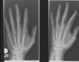 the human hand with it's opposable
thumb that gives us the advantage over even the apes and chimps that anthropologists say
are our closest cousins.Ā With 27 bones making up the hand and wrist, it also one of
the most complex part of the human body with more ways existing to x-ray it than perhaps
any other part of the body except the head.Ā As a result, hands and wrist normally
require at least three pictures, and sometimes four or more, to adequately demonstrate all
the different parts.Ā Since the main difference between the three most common x-ray
positions for the hand and the wrist are where the x-rays are centered, I will deal with
both of them together and then detail the various specialty views of each separately. the human hand with it's opposable
thumb that gives us the advantage over even the apes and chimps that anthropologists say
are our closest cousins.Ā With 27 bones making up the hand and wrist, it also one of
the most complex part of the human body with more ways existing to x-ray it than perhaps
any other part of the body except the head.Ā As a result, hands and wrist normally
require at least three pictures, and sometimes four or more, to adequately demonstrate all
the different parts.Ā Since the main difference between the three most common x-ray
positions for the hand and the wrist are where the x-rays are centered, I will deal with
both of them together and then detail the various specialty views of each separately.Whether you have an injury to your hand or your wrist, three
main views will be done that differ between hands and wrist only in whether the x-rays are
going to be centered to the hand or the wrist, and in the position of the fingers.Ā
For each you will have an x-ray with the hand and wrist flat against the film, a side or
One reason that three views of the wrist are needed to adequately x-ray it is that the bones that make up the majority of the wrist, called carpals, form a loose C if view looking down along the arm.Ā As a result, the bones at the base of the thumb (and the thumb itself) are only seen in a flat, frontal view when the hand is rotated up to a 45 degree angle.Ā Yet this ignores the the carpal bone at the other end of the "C", called the pisiform.Ā The pisiform is one of, if not the smallest bone in the body, and as such, it is extremely difficult to break. Ā This is why many facilities don't bother trying to get a better picture of it than can be seen on the PA, or frontal view.Ā However, extremely difficult to break is not the same as impossible to break, and if the doctor thinks that you may have broken it, there is a forth view of the wrist that can be done to show the pisiform in full detail. Ā This position is another oblique, or angle shot, this time called a semisupanation oblique (pronounced semi-soupanation).Ā As in the semipronation view, the hand forms a 45 degree angle with the film, with the pinky against the film and the thumb up in the air.Ā But, instead of the palm facing towards the film in this view, it is the back of the hand that is turned towards the film.Ā This leave the pisiform sitting out away from the other bones in the hand and wrist on the x-ray, looking for all the world like a small little lost pea. Another carpal bone that we occasionally need a special picture of is the Scaphoid or Navicular (either word is correct though scaphoid is the newer "preferred" name) which sits at the base of the thumb.Ā Since it is this bone that helps form the base that makes our thumb an opposable thumb and allows us to grip things better, the bone itself sits at a double angle to the rest of the wrist. Ā Again, like the pisiform, the navicular is a very difficult bone to break, but it can be done, and when it is fractured it requires special consideration if the doctor is to be sure that your thumb will continue to work correctly.Ā It doesn't have to be perfect, but if it is too badly deformed, then you will have difficulty gripping things tightly and firmly for the rest of your life.Ā To x-ray the navicular, the technologist will have you twist your hand so that while flat, the fingers will point away from your body.Ā A small wedge shaped sponge or a folded towel will then be placed under your hand and the film so that the film runs up hill at about a 20 degree angle. Ā This will separate the navicular or scaphoid from the rest of the bones of the wrist and allow us to view dead on without any other bones being superimposed on top of one end or the other. At the other end of the hand is, of course, the fingers and thumb. Ā The fingers themselves are normally x-rayed using the same positions as the hand except that only one finger is extended so that the finger in question is the only one seen on all three pictures.Ā The only minor difference is that in the side or lateral view, the hand is rotated in which ever direction will put the injured finger closest to the film in order to avoid making the finger look larger than it really is.Ā The thumb however takes a bit of twisting of the wrist to correctly x-ray.Ā This is because the thumb is turned 45 degrees from the plain the rest of the hand lies in. Ā As a result, what would provide a frontal shot of the rest of the hand actually gives us an angled shot of the thumb.Ā Indeed you can see this for yourself just by looking at your own thumb.Ā When you rest your hand flat, the thumb nail is at a 45 degree angle to the surface you are resting your hand on.Ā Twist your wrist so that your hand is now at a 45 degree angle to the surface you are resting your hand on, and the thumb nail is now perpendicular to that surface, providing us with a perfect side shot.Ā The tricky one is the frontal shot though.Ā Slide your hand to the edge of the table or desk you are sitting at, and you can place your thumb flat on the desk, but the portion of your wrist that makes up the base of the thumb is now hanging off in space.Ā Now twist your wrist so that you can place the back of your thumb flat on the desk, and the entire thumb from the tip of the thumb nail to the wrist is now flat against the desk.Ā This is the position that we must use if the entire thumb is to be shown on the x-rays.Ā Admittedly it is a bit uncomfortable, but if you use mainly motions of the shoulder and elbow, then even if your thumb is broken we can still achieve a good frontal (or in this case, back) view of the thumb. There are a few more specialized x-rays we can use for the hand and
wrist, but for the most part these are rarely used except in certain circumstances, and
thus are beyond the subject of this page.Ā If you wish information about any of these
special views, e-mail me, Carl Kurtz, and I will
do my best to provide what information I can, or call the facility at which your exams
will be done. |
[Home]ĀĀ [Cat Scan]ĀĀ [MRI]ĀĀ [Mammography] ĀĀ [Sign Guestbook]ĀĀ [Bio] |
|
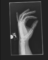 lateral view with the pinky side of the hand and wrist against the film and the thumb side
facing the x-ray tube, and a semipronated oblique with the pinky side of the hand
and wrist against the film and the thumb side up in the air so that the palm faces the
film and the hand forms a 45 degree angle with the film.Ā the only real difference in
these views between those done for a hand and those done for a wrist is, as I said
earlier, in the way the fingers are held.Ā For the PA view (hand and wrist flat
against the film) the fingers are as flat as possible and slightly fanned out for a hand
x-ray, while for a wrist the fingers are curled under into a loose fist to help press the
wrist closer to the film if possible (sometimes the fingers can not be curled for one
reason or another, but the lose of film quality this causes is not enough to make any real
difference most of the time).Ā The other picture where there is a difference in
finger position between a hand x-ray and a wrist x-ray is in the side or lateral
view.Ā When doing a hand x-ray, the technologist will ask you to try to fan the
fingers into something similar to an "okay" sign so that each finger will be
visible on the x-ray, while in a wrist picture it really doesn't matter what position the
fingers are in though some technologist feel that the patient is less likely to
accidentally move their hand out of position if the fingers are again curled into a loose
fist.
lateral view with the pinky side of the hand and wrist against the film and the thumb side
facing the x-ray tube, and a semipronated oblique with the pinky side of the hand
and wrist against the film and the thumb side up in the air so that the palm faces the
film and the hand forms a 45 degree angle with the film.Ā the only real difference in
these views between those done for a hand and those done for a wrist is, as I said
earlier, in the way the fingers are held.Ā For the PA view (hand and wrist flat
against the film) the fingers are as flat as possible and slightly fanned out for a hand
x-ray, while for a wrist the fingers are curled under into a loose fist to help press the
wrist closer to the film if possible (sometimes the fingers can not be curled for one
reason or another, but the lose of film quality this causes is not enough to make any real
difference most of the time).Ā The other picture where there is a difference in
finger position between a hand x-ray and a wrist x-ray is in the side or lateral
view.Ā When doing a hand x-ray, the technologist will ask you to try to fan the
fingers into something similar to an "okay" sign so that each finger will be
visible on the x-ray, while in a wrist picture it really doesn't matter what position the
fingers are in though some technologist feel that the patient is less likely to
accidentally move their hand out of position if the fingers are again curled into a loose
fist.