| Ā | Ā Head & Sinus X-rays Ā |
| Skull Films
Special X-ray Units for Head Films Ā Ā Ā
|
Ā Head, or Skull, and Sinus x-rays are To help overcome some of these difficulties, two basic types of x-ray devises have been developed over the years, along with a host of different types of holders designed to help get and keep the head into position. The first of these devises is the Franklin Head Unit, an odd looking devise mounted on a thick metal post or column that reaches from the floor to the ceiling. Ā The unit includes a metal film holder with a glass or plastic front and a hole in the back in the center of the holder, plus an x-ray tube mounted on a arm that swings up and down, so that the x-ray tube moves in an arc about the film holder.Ā The patient is placed in a specially designed chair that can be move about freely until the patient is in the right position, then locked down securely so that it will not move again until the locks are released.Ā The hole in the back of the film holder is placed in such a way that the technologist doing the study can look through the hole to see just how the patient is positioned before the film and any filters, or grids, are inserted into the holder.Ā The unit also commonly has a pair of padded arms that can be used to help hold the patients head in place once the technologist has him or her in the proper position. There are two big drawbacks to the Franklin Head Unit though.Ā First is the fact that it can only be used to do head x-rays.Ā Since some of the exams that this unit really shines on, such as Mastoids and TMJ's (short for Temporal Mandibular Joints which refers to the point where your jaw meets the skull) are now more commonly done in Cat Scan, many facilities do not feel the expense of such a unit is justifiable.Ā The second big drawback is that for one reason or another, this unit is the type of thing that you either love or hate.Ā I don't know any technologists who fall in the middle. The second class of devises are variations of the Panorex unit.Ā A panorex uses a curved x-ray plate and a special unit that moves the x-ray tube around your head in a motion similar to a Tomography unit (see the IVP page for an explanation of tomographs).Ā Some units require the patient to sit or stand, and some units have the patient lay on a specially design table, but either way the patient's head is placed in position using special shields as guides and then generally secured in place.Ā The machine will then be turned on, causing the curved x-ray plate to pass around in front of the patient's head while the x-ray tube, or camera passes around in back.Ā The whole devise tends to look like something out of a science fiction movie, and many people find the motion of the x-ray plate and tube slight unnerving.Ā There is no cause for alarm however as nothing will touch you once the machine is turned on until the exam is over. Ā The big reason for using this devise is that it turns out x-rays that have the appearance of someone having peeled the bones being studied off and flattened them out just like a label off a bottle.Ā The two most common exams this type of machine is used for are jaws, or mandibles, and teeth. When you report to the radiology department for your head x-rays, no matter what type that you are getting, the first thing the technologist in charge of your study will do is have you remove any metal items from your hair, ears, neck, etc.Ā These items include such things as: hair pins, ear rings, glasses, necklaces, dentures and partials, and of course for the younger and/or more "hip" crowd, nose rings, lips rings, etc.Ā Depending on what kind of shirt or blouse you are wearing, you may also be asked to change into a hospital gown as an added precaution.Ā You will then be escorted to the x-ray room where your exam will be done. Skull, or Cranium films generally take three to five films to complete depending on the facility you are having your x-rays done at and what your doctor thinks may be wrong.Ā These pictures are commonly done upright when possible as some skull fractures are more easily spotted by the blood or other fluids that drain from the fractures into the sinuses and air cells in the skull.Ā The first picture or two will almost always be done with you facing towards the film holder, and the last x-ray will be done with your back to it. A side shot, called a lateral will definitely be included in the series as well. Facial x-rays are also commonly done upright, and for the
same reason as skull films.Ā Sometimes the physician examining you will be more worried about the eye sockets, or orbits than the rest of the face, in which case he or she may order x-rays focused on just these bones.Ā Once again the films will be done with you, the patient in as upright a position as possible to make any blood or fluid draining into the sinuses around the eyes more easily seen since this is once again a good sign that fractures that are not otherwise evident do indeed exist.Ā The basic orbit series consists of three films centered around the eyes.Ā Two with you facing the film holder is possible, and a side shot.Ā From there however it gets tricky.Ā Some facilities automatically add in additional specialty views on all patients just as a precaution, while others only use these additional views if the injuries involved suggest that they may be needed. Ā One hospital I once worked at went so far as to use a device similar to the panorex machine described above to get tomographic like "slices" of the bones around the eyes to check for fractures that did not go all the the way through the bone (for a discussion of tomography, please see the IVP page on this site). Another subset of facial films are x-rays of the sinuses.Ā These films almost have to be done upright since they are most commonly done to check for sinusitis (an infection in or of the sinuses) or similar problems, which are normally indicated by fluid in the sinuses.Ā If the patient is laying down, any fluid in the sinuses will cover the entire back wall of the sinus involved making it difficult to see, and even harder to measure.Ā The three sets of sinuses that the technologist will be most interested in capturing on x-rays are: the frontal sinuses which are located between and directly above the eyes and look something like broccoli, the maxillary sinuses which look like small triangles directly below the eyes, and the sphenoid sinuses which are located directly behind the eyes.Ā Typically four views or pictures will be used to see these sinuses, two with you facing the film holder to demonstrate the frontal and maxillary sinuses, a picture with you head tilted as far back as you can and shot up through the chin and rood of you mouth to see the sphenoid sinus, and a side shot, or lateral to see all three from the side.Ā Some facilities will have the technologist use a cone, which is a long hollow tube that attaches to the front of the x-ray "camera" to better demonstrate the sinuses.Ā If the facility at which you are having your x-rays done uses this device, I strongly urge you to be sure not to move your head during the x-rays until the technologist specifically tells you to .Ā You see, the cone will come to within 18 inches of the back of your head during the x-rays, and if you suddenly move your head too far, you will end up with a major headache. The final set of head x-rays I will talk about here are of the mandible, or jaw bone.Ā This is probably the hardest part of the head to x-ray, since the jaw bone looks something like a cross between a hastily drawn U and a rounded V. Ā Traditionally, a mandible series consists of five x-rays, though some facilities will use a panorex type machine instead.Ā The five x-rays traditionally used are: a sharply angled front shot (called a Low Townes) to check for side to side alignment of the bones, a straight side shot or lateral to check for up and down alignment of the bones, a straight on front shot to look for fractures in the chin area, and two obliques or angled shots from the sides to look for fractures on the sides of the jaw.Ā It is these two obliques that often cause the technologists the most problems.Ā The trick is that each side must be seen without having the other side superimposed over it, which means tilting your head and angling the x-ray tube.Ā It is also important to see the section of your jaw bone that connects with you head without the spine showing up in the middle of everything.Ā This means that the patient must also turn his or her head slightly to the side as well as tilting it.Ā All of which tends to conspire to place the shoulder smack dab in the middle of everything, especially if you have big shoulders. Ā As a result, it is not uncommon for even the most experienced technologist to take two, three, or even four tries at it before he or she gets a usable film.Ā This is one reason why many facilities have taken to using panorex to x-ray the jaw even though the panorex will often distort the chin portion of the image. Finally, one word of warning about asking your technologist what your x-rays show.
Ā Since the technologist does not have the training to read the x-rays the way a
medical doctor does, and federal law severely frowns on them even trying, most
technologists will not tell you anything about any of your x-rays anyway.Ā But with
any type of head x-rays, the difficulty in reading the films is compounded to the point
where even those technologist who will give you some information probably won't even try.
Ā So please.Ā Save yourself and the technologist some aggravation, and don't
even ask. |
[Home]ĀĀ [Cat Scan]Ā [MRI]Ā [Mammography]Ā [Sign Guestbook] ĀĀ [Bio] |
|
 commonly among the last plain film x-rays
taught in school, and for the simple reason that they can be the hardest to do.Ā To
begin with, the skull is not just one bone, but rather is made up of 22 different bones
that, with one exception, join together at immovable joints called sutures.Ā
Then there is the overall shape of the head.Ā It's not exactly round, or spherical,
like a ball, nor is it truly egg shaped, or any other shape.Ā It is only Head
shaped.Ā Then there is the fact that preferably you should do most of the films of
the head with the patient facing the film holder, or bucky, to avoid making the eye
sockets, called orbits, appear larger than they really are.Ā Couple all of
this with a healthy head of hair, and you have a body part that is very difficult to
position properly, especially since you can't trust your eyes to tell you when the
patient's head is straight.
commonly among the last plain film x-rays
taught in school, and for the simple reason that they can be the hardest to do.Ā To
begin with, the skull is not just one bone, but rather is made up of 22 different bones
that, with one exception, join together at immovable joints called sutures.Ā
Then there is the overall shape of the head.Ā It's not exactly round, or spherical,
like a ball, nor is it truly egg shaped, or any other shape.Ā It is only Head
shaped.Ā Then there is the fact that preferably you should do most of the films of
the head with the patient facing the film holder, or bucky, to avoid making the eye
sockets, called orbits, appear larger than they really are.Ā Couple all of
this with a healthy head of hair, and you have a body part that is very difficult to
position properly, especially since you can't trust your eyes to tell you when the
patient's head is straight.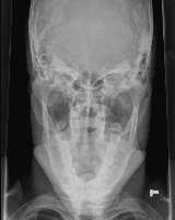 A facial series of x-rays often use the same views as
skull films except since the face is the area of interest instead of the the cranium, or
brain case, the films are normally centered lower and done at a lighter setting.Ā
However there are a couple of views often included in facial x-rays that will not be used
for a routine skull series.Ā Which of these additional views are used will depend on
what your main problem, or chief complaint is.Ā For example, if you were assaulted
and the doctor is worried that your cheek bone (called the zygoma or zygomatic
arch) is broken, a set of x-rays called jug handles may be included.Ā
In this picture, the technologist will set things up so that the x-ray plate is against
the top of your head and the x-ray tube will be shooting up from under your chin.Ā
The resulting image, called an axial view, will show the cheek bones sticking out
from the side of your head like a set of handles on a decorative jug, thus the common nick
name.
A facial series of x-rays often use the same views as
skull films except since the face is the area of interest instead of the the cranium, or
brain case, the films are normally centered lower and done at a lighter setting.Ā
However there are a couple of views often included in facial x-rays that will not be used
for a routine skull series.Ā Which of these additional views are used will depend on
what your main problem, or chief complaint is.Ā For example, if you were assaulted
and the doctor is worried that your cheek bone (called the zygoma or zygomatic
arch) is broken, a set of x-rays called jug handles may be included.Ā
In this picture, the technologist will set things up so that the x-ray plate is against
the top of your head and the x-ray tube will be shooting up from under your chin.Ā
The resulting image, called an axial view, will show the cheek bones sticking out
from the side of your head like a set of handles on a decorative jug, thus the common nick
name.