| Ā | Ā Legs |
| Ā
Standing or Wieght Bearing Films Ā Ā This page hosted by
Get your ownFree Home Page
|
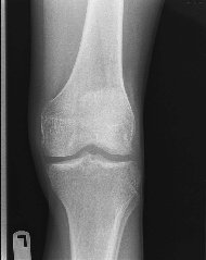
Ā Though we tend to take them for The biggest and strongest bone in the human body is the Femur, which is the bone in the upper leg or thigh.Ā Looking for all the world like a Caveman's answer to the heavy war hammers of the middle ages, the femur is a large, straight bone with a ball shaped head that sits at an angle to the shaft of the bone.Ā Generally only two views of the femur are needed, a frontal view or AP, and a side shot called a lateral. Ā However don't expect the technologist doing your x-rays to get your entire femur with only two pictures.Ā For one thing, the average adult has a femur that, according to one anatomy book I've read, is 19 to 21 inches (that's 48 to 53 centimeters) in length while the largest x-ray film that is not designed for a special purpose is only 14 inches x 17 inches (or 35 cm x 43 cm).Ā Then to make matters worse, since the head of the femur connects to the hip, even on a short person or child two films will be needed to show the entire femur from the side.Ā The AP view is easy to do, the technologist will simply take one film centered to get the top of the femur to a point as close to the knee as possible, and then a second pictureĀ centered to get the knee to a point as close to the hip as possible.Ā However the lateral view of the femur is a bit tricky. Ā The clearest x-rays are obtained by bending the knee of the affected leg up and then letting the leg fall out as far to the side as possible.Ā Two films can then be done centering each film the same way the AP shots are centered.Ā Unfortunately this is often not possible when the patient comes into the emergency room, especially if the femur, the knee, or the hip is fractured.Ā In cases like this, it is often necessary to use what are commonly called cross table laterals, where the x-ray camera is turned sideways so that the x-ray beam is parallel to the floor.Ā While the film showing the lower portion of the femur can be done by placing the x-ray cassette between the patient's legs, this will not show the top portion of the femur where it connects to the hip.Ā To get a side view of this section of the femur, it is necessary to place the film beside the hip on the injured side, raise the uninjured leg as high up in the air as possible, and shot through the opening thus created. The Knee is made up of the junction of four bones, namely the Femur, the Tibia, the Fibula, and the Patella or Knee Cap. Ā Depending upon which facility you have your x-rays done at and what your problem is, a knee series will consist of between two and four x-rays.Ā The first x-ray will be a frontal or AP shot where the film will be placed behind the patient's knee and the x-ray camera will be angled slightly towards the patient's head.Ā This helps to open up the joint between the femur and the Tibia and fibula thus allowing us to see if anything has gotten caught in this space.Ā For the side shot, or lateral, the patient is commonly rolled up onto the side of interest with the upper leg brought forward so that it is out of the way of the affected knee.Ā The knee we are x-raying will then be bent at a slight angle which helps to keep the knee in a true lateral position.Ā Two additional views that may be included depending upon where you go and what is wrong are two obliques or angled shots.Ā For these two x-rays, the patient is asked to roll his or her leg about 45 degrees outwards, then in towards the other leg.Ā This allows the doctor to evaluate the joint between the fibula and the tibia, the patella, and the femoral condyles, which are a pair of knurled, rounded knots on the end of the femur which helps to allow the knee to bend the way it does. Occasionally your doctor may be concerned with the Patella, or Knee Cap itself.Ā It could be that he or she is worried that the patella itself is fracture, or your doctor may just suspect that it is misaligned or dislocated.Ā What ever the reason maybe, the technologist will be called upon to do an x-ray commonly called a Sunrise View, so called because it demonstrated the knee cap floating above the knee itself just like the sun over the ocean at sunrise.Ā To do the x-ray, the patient can be either sitting or laying on his or her stomach.Ā The patient is then asked to bend his or her knee to as close to 45 degrees as he or she can, and a film is placed on the patient's thigh so that when the x-ray is shot from near the patient's toes, the knee cap will be close to the center of the film.
Sometimes you doctor may be concerned about a variety of problems that will cause your legs and/or knees to shift in an abnormal manner whenever you stand or walk.Ā In this case he or she may order a set of weight-bearing films which can be of the knees, the hips, or the entire leg.Ā No matter which of the three it is however, AP's and Lateral similar to those discussed about will be shot of both legs while the patient is standing upright in order to evaluate any deformities that may show up. |
[Home]Ā [X-rays]Ā [Cat Scan]Ā [MRI]Ā [Mammography]Ā [Sign Guestbook]Ā [Bio] |
|
 granted, our legs are one of the more complex
parts of the body; not in terms of design but rather in the number of things that can go
wrong.Ā Yet inspite of this, x-rays of the legs is fairly straight forward without
even those little things that trip technologists up on other parts of the body such as
arms and skulls.
granted, our legs are one of the more complex
parts of the body; not in terms of design but rather in the number of things that can go
wrong.Ā Yet inspite of this, x-rays of the legs is fairly straight forward without
even those little things that trip technologists up on other parts of the body such as
arms and skulls.