
Sign The Guestbook!
Read The Guestbook!
since Feb 26, 1998
This page hosted by
Get your own
Free Home Page

|

Sign The Guestbook! Read The Guestbook! since Feb 26, 1998 This page hosted by Get your own Free Home Page |
|
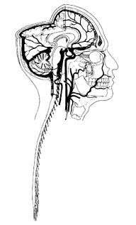
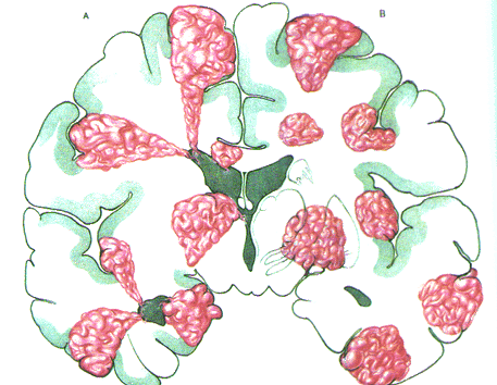
Arteriovenous Malformations (AVMs) Arteriovenous Malformations (AVMs) are abnormal, tumor like masses of vessels in the brain or spinal cord. Normally, the arteries entering the brain or spinal cord divide continuously into smaller branches until they reach the stage of the capillaries. Through the capillaries, oxygen and glucose are delivered to the brain tissue and the useless end products of its metabolism are removed. The capillaries continue into very small veins, which continuously unite to form larger ones and finally conduct the blood out of the brain or spinal cord. The capillary system also serves another purpose, to lower the high pressure of blood in the arterial system so that the venus system will have a lower one which it can tolerate. Arteriovenous malformations occur when the capillaries are absent and there is direct communication between the arterial and the venous side of the circulation. This means that there is direct transfer of high pressure blood from arteries into veins. Veins cannot whithstand such a high pressure and they are forced to dilate and elongate in order to accomodate it. Following the dilatation and elongation of the veins, a mass of vessels is formed which is called an arteriovenous malformation. Below are some drawings of various types of AVMs: 

 
 LEFT: Various locations of AVMs in the brain
RIGHT: Drawing of AVM in the brain Below are images from MRI scans of an AVM: 
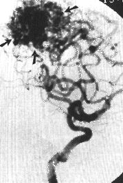
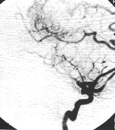
 RIGHT: Sagittal MRI scan An arteriovenous malformation can be harmful to the brain or the spinal cord in two ways: 1. By direct pressure on the sensitive brain or spinal cord tissue. Depending on the exact location of the AVM and the amount of pressure it causes, various kinds and degrees of neurological deficits can occur. Pressure on some areas of the brain can even cause epileptic seizures. 2. By hemorrhage caused by rupture of the veins due to the high blood pressure. This can be a very serious and even life thratening condition or it can be a milder one depending on the severity of the bleeding and the location in which it occured. Neurological deficits produced by a hemorrhage can be permanent or improve gradually with time. The cause for the formation of AVMs is not yet known. It is believed to be some genetic mistake governing the development of blood vessels. AVMs can be present at birth or can develop later in life. There are currently three treatment options for AVMs: 1. Conventional surgery. The neurosurgeon performs a craniotomy to enter the brain, reach the AVM and remove it. It offers very good results but can have a significant rate of complications due to the operative procedure itself or to the fact that an AVM might not be easily separated from normal tissue. There are AVMs located deep into the brain or the spinal cord which are considered inoperable. 2. Radiosurgery. It is performed with a gamma-knife or an x-knife, radiotherapy devices which can target the radiotherapy beams directly to the AVM relatively sparing healthy tissue causing to it less damage than conventional radiotherapy. Their effectiveness is limited by the size of the AVM (up to 2 cm in diameter) and by the fact that it takes up to 2 years for the effect of radiation on the AVM to take place following treatment. This means that for a period of up to 2 years an AVM can still cause pressure on the brain, epileptic seizures or bleedings. 3. Endovascular neurosurgery. The AVM is treated by reaching it with very fine catheters through a puncture of the femoral artery under x-ray control and injecting into it materials which block its vessels and stop blood from entering it. The effect on the risk of bleeding is immediate and the AVM gets smaller until it disappears in 6-12 months. Most inoperable AVMs can be treated with this method with excellent results. There are almost no associated risks and the hospitalization and recovery periods are very short. Below is an example of an AVM treated with the method of Endovascular Neurosurgery: A  B
B  A. Angiogram of a brain AVM B. AVM treated with Endovascular Neurosurgery method Page maintained by: Vasilis Katsaridis, MD |