
Sign The Guestbook!
Read The Guestbook!
since Feb 26, 1998
This page hosted by
Get your own
Free Home Page
|
|
|
Sign The Guestbook! Read The Guestbook! since Feb 26, 1998 This page hosted by Get your own Free Home Page |
|
Brain Tumors The same techniques can be applied with the same advantages in tumors of the face and neck as well. The most common of these tumors are the juvenile nasopharyngeal angiofibromas, benign tumors of the nasopharynx which can bleed dangerously during an operation. Following embolization they can be removed with minimal blood loss. Below are some angiographic examples of brain tumors embolised with PVA particles with the use of Endovascular Neurosurgery techniques: Case 1 A  B B  A: Angiographic appearance of a frontal meningioma B: Same tumor following embolization with PVA particles Case 2 A 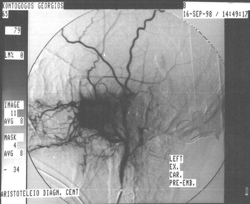 B B 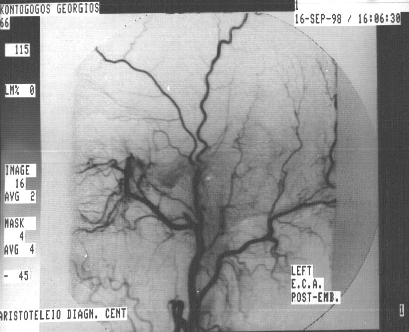 A: Angiographic appearance of a skull base meningioma B: Same tumor following embolization with PVA particles Case 3 A  B B  A: Angiographic appearance of a jugular foramen paraganglioma (glomus jugulare tumor) B: Same tumor following embolization with PVA particles Page maintained by: Vasilis Katsaridis, MD |