Structural comparisons between fungal peroxidases and mammalian peroxidases
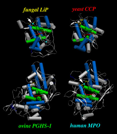
Fig.5 Structural comparisons between fungal peroxidases and mammalian peroxidases suggest that they arose from a common percursor. Here, the structures of lignin peroxidase (LiP), cytochrome c peroxidase (CCP), myeloperoxidase (MPO), and PGHS peroxidase share the same features such as catalytic site helices (green), the scaffolding helices (light blue) around the heme (red) and functional (arrows) or vestigial (*) calcium binding sites.(6)

Fig.6 Accessible surface area views of the fungal peroxidases and mammalian peroxidases show how buried the heme (red) is in many peroxidases except PGHS, which uses larger hydrophobic hydroperoxides (e.g., PGG2, 15-HPETE, etc.) as substrates rather than hydrogen peroxide. The structures shown are lignin peroxidase (LiP), cytochrome c peroxidase (CCP), myeloperoxidase (MPO), and PGHS peroxidase.(6)