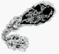
The Circulating form of dendritic cell precursor looks like a lymphocyte with an irregular nucleus and numerous mitochondria in the cytoplasm. Click image to see LY-DC comparison.

Dendritic Cells In Tissue

I was curious why the dendritic cells in Thoracic Duct Lymph [TDL] were not obvious, so I decided to try and get a look at them by EM. I did "panning" experiments to isolate the "precursor" of DC in TDL. B- and T cells were removed by panning on cell-specific antibody coated plates. I removed phagocytic cells by short term incubation and/or particle phagocytosis. I selected cells with high expression of Class II MHC for EM. The most prevalant morphology of these cells (presumptive DC) is at the left. This "DC-precursor" found in TDL looks very much like a lymphocyte with several important exceptions. Mitochondria were far more numerous in the cytoplasm, and the DC nucleus was convoluted with more delicately distributed heterochromatin and lighter euchromatin than is normally found in lymphocytes. For comparison, Look at the nucleus of the lymphocyte at the bottom of the figure below.

This mature Interdigititating Dendritic Cell is in contact with the fluid conducting fiber of the FRCC. Note that no cell cytoplasm or membranes separate the DC from the dark fiber. The fiber probably retains contact as it passes behind the IDC. Contact with the fiber may permit the IDC to sample chemokine signals and/or antigens flowing in the conduit.
As the dendritic cell precursor begins to differentiate it develops this tadpole shape
Upon attachment to the fibroblastic reticular cell conduits in lymphatic tissues, the dendritic cell asssures that its membrane is in contact with the fluid environment of the conduit at at least one point. Note the DC process making contact with the FRCC fiber between apposed membranes of the ensheathing reticular cell. Click for more detail.

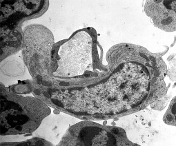
The two tadpole shaped cells above are probably intermediate forms in dendritic cell maturation, (commentary). The upper image was found in lumen of a blood vessel from an immunized rat 7 days after it survived infectious challenge. Dendritic cells at this stage of maturation would probably not be found circulating. The lower cell just above this text is very similar in morphology (ie tadpole shaped) with the exception that it is anchored to a fibroblastic reticular cell conduit (FRCC, seen as a cross section). It is clear from EM and immuno- histochemistry studies that DC anchored to FRCC participate in efficient acquisition and presentation of antigens to lymphocytes that continuously emigrate from the blood across HEV and enter the channels, corridors and cords of the the lymph node cortex. Dendritic cells are found distributed in the LN cortex near HEV but they also may be found in the walls of HEV, especially when immune stimuli triggers inflammation.

For information on factors influencing dendritic cell development and differentiation, see Immunol. Rev. 156:25.

Click HERE to return to FRCC.
Click HERE to return to Homing.

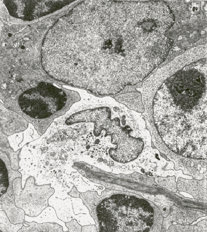
The EM above is from the deep cortex of a regional lymph node of a rat 48 hours after application of an allogeneic skin graft. Within the field of view you can see a macrophage (large cell at top) an interdigitating dendritic cell (light cell in the middle attached to an FRCC, crossing diagonally from lower right), and a T lymphoblast (cell with large nucleus and nucleolus, middle right). One might imagine that this scene is where graft alloantigens are being efficiently presented to the numerous (up to 200) cells with whom the IDC processes may synapse [PubMed].

  • Steinman, R.M., 1991. The dendritic cell system and its role in immunogenicity. Annu. Rev. Immunol. 9:271-296.
  • Vremec,D., G.J. Lieschke, A.R. Dunn, L. Robb, D. Metcalf and K. Shortman. 1997. The influence of granulocyte/macrophage colony-stimulating factor on dendritic cell levels in mouse lymphoid organs. Eur. J. Immunol. 7:40-44.
  • Steinman, R.M. and J. Swanson. 1995. The Endocytic Activity of Dendritic Cells. (Commentary) J. Exp. Med. 182:283-288.
  • Macpherson, G.G. 1989. Properties of lymph-borne (veiled) dendritic cells in culture. I. Modulation of Phenotype, Survival and Function: Partial Dependence on GM-CSF. Immunol. 68:102-107.
  • Fossum, S. 1988. Lymph-Borne Dendritic Leukocytes do Not Recirculate, but Enter the Lymph Node Paracortex to Become Interdigitating Cells. Scand. J. Immunol. 27:97-105.
  • Bell, E.B. and J. Botham. 1982. Antigen transport I. Demonstration and Characterization of Cells Laden With Antigen In Thoracic Duct Lymph and Blood. Immunol. 47:477-487.
  • Peters, J.H., R. Gieseler, B. Thiele, and F. Steinbach. 1996. Dendritic Cells: from ontogenetic orphans to myelomonocytic descendants. Immunol. Today 17:273-277.

  • ArtnScience


    Recirculation | Emigration at HEV | FRC Conduit | Locomotion | Chemotaxis & Links

    Dendritic Cells in TDL | Dendritic cells in LN Tissue | Mucosal and Peripheral Immunity

    Immunology Lecture Notes | Panama Canal Builder | Art's Curriculum Vitae

    StatNislander's Photo Album | The Army and BioEthics | Art's Art 'n Stuff


    Counter Reset 15 April 1999