Approaches to Interpretation of CT
![]()
Head
| Prior imaging | oldest & most recent PET, MRI |
| Scout | Lines, tubes, #'s, C-spine |
| Bone window | #'s, sinuses, lesions |
| Brain window | mass effect, blood (esp. SDH), edema, gray-white differentiation, CSF, arteries (aneurysms, calcifications) |
| Soft tissue window | Vessels, LN's, salivary glands, thyroid |
-
Chronicity of Hemorrhage
| 0 - 2 hrs | 40 - 60 HU |
| 3 - 48 hrs | 60 - 80 HU |
| 3 - 7 days | hyperdense core surrounded by hypodense halo |
| > 2 wks | hypodense |
Edema
Vasogenic:
Due to increased permeability of brain capillary endothelial cells to plasma proteins. Greatest in white matter. Astrocytes become swollen.
E.g. trauma, tumors, focal inflammation, and late stages of cerebral ischemia.
Cytotoxic:
Characterized by swelling of all the cellular elements of the brain. In the presence of acute cerebral ischemia, neurons, glia, and endothelial cells swell within minutes due to failure of ATP-dependent ion (sodium and calcium) transport. With the rapid accumulation of sodium within cells, water follows to maintain osmotic equilibrium. Increased intracellular calcium activates phospholipases and the release of arachidonic acid, leading to the release of oxygen-derived free radicals, and infarction.
E.g. various intoxications (dinitrophenol, triethyltin, hexachlorophene, isoniazid), Reye's syndrome, severe hypothermia, and early ischemia.
![]()
Spine
Critical spinal stenosis, AP dimension of thecal sac:
1.0 cm
1.4 cm in L-spine
![]()
Chest
| Prior imaging | oldest & most recent PET, MRI |
| Scout | Lines, tubes |
| Lung window | Airways: endobronch lesns bronchial wall thickening (wall thickness > diameter adjacent vessels) bronchiectasis (cylindrical, varicose, cystic)
Lung parenchyma: |
| Soft tissue window | Flow of contrast, heart, great vessels, LN's Abd (ascites, free air, vessels, adrenals, kidneys, spleen, pancreas) |
| Liver window | Liver (density, lesions, cirrhosis) |
| Bone window | Lines, #'s, lesions, invasn |
| Normal | Else
| Main PA < 3 cm
| Indicative of PA hypertension
| Aorta: | Root < 3.6 cm Ascending < 3.5 cm Proximal descending < 2.6 cm Distal descending < 2.4 cm Abd < 3 cm Aneurysm | If < 4 cm → ectasia If > 4 cm → dilatation
|
|
|
| |
Density > 200 HU indicates calcification
Cavity wall thickness:
< 2 mm, 95% benign
2 - 15 mm, 50% malignant
> 15 mm, > 95% malignant
Eccentric cavity or shaggy internal margins suggests malignancy.

Regional Nodal Stations for Lung Cancer Staging
(American Joint Committee on Cancer(AJCC) and the Union Internationale Contre le Cancer (UICC))
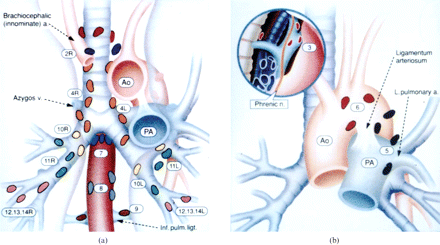
1 (red) = highest mediastinal nodes
2R and 2L (dark blue) = right and left upper paratracheal nodes
4R and 4L (orange) = right and left lower paratracheal nodes
7 (blue) = subcarinal nodes
8 (grey) = para-oesophageal nodes
9 (brown) = pulmonary ligament nodes
10R and 10L (yellow) = right and left hilar nodes
11R and 11L (green) = right and left interlobar nodes
12R and 12L (pink) = right and left lobar nodes
13R and 13L (pink) = right and left segmental nodes
14R and 14L (pink) = right and left subsegmental nodes
3 (pink) = pre-vascular and retrotracheal nodes
5 (black) = subaortic nodes
6 (red) = para-aortic nodes
Fleischner Society Recommendations for Incidental Pulmonary Nodule Follow-up
Multinodular Disease: A High-Resolution CT Scan Diagnostic Algorithm
Abdomen
| Prior imaging | oldest & most recent PET, MRI |
| Scout | Lines, tubes |
| Liver window | Density, lesions, cirrhosis |
| Soft tissue window | Skin, hernia,
adrenals, kidneys, spleen, pancreas,gallbladder, LN's ureters, bladder, bowel, terminal ileum, appendix prostate, seminal vesicles, uterus, ovaries |
| Bone window | #'s, lesions |
| Lung window | Lung bases |
-
Liver lesions
| Cyst | Thin, smooth wall 0 - 15 HU No enhancement No septations |
| Cavernous hemangioma | Discontinuous, nodular peripheral enhancement Centripetally-filling Isodense to aorta |
| Adenoma | Heterogen enhancement Well-defined capsule |
| FNH (fibrolamellar HCC similar) | Art phase: homogen enhancement (key) Solid (isodense when unenhanced) Central scar Radiating fibrous septa Portal venous phase: ↑ periph enhancement (dt lge pariph veins [no capsule]) |
Bosniak classification of cystic renal masses by CT scanning
| Category | Meaning | Criteria
| I
| Simple benign cyst
| Hairline thin wall
| Density less than 20 HU(similar to water) No septa, calcification, or solid components No enhancement II
| Benign cyst
| A few thin ( < 1 mm thick) septa
| No measurable enhancement (may be "perceived" enhancement) Includes uniformly high attenuation lesions <3 cm that are well marginated and do not enhance IIF
| Minimally complicated cyst that requires follow-up
| Multiple hairline thin septa or minimal smooth thickening of the wall or septa
| No measurable enhancement (may be "perceived" enhancement) Thick and nodular calcification of the wall or septa Totally intrarenal, nonenhancing, high attenuation lesions >3 cm III
| True indeterminate cystic mass that typically undergoes surgical evaluation
| Thickened wall or septa in which measurable enhancement is present
| IV
| Mostly malignant
| Enhancing soft-tissue components adjacent to, but independent of, wall or septum
| |
N.B. Enhancement = attenuation increase by at least 10 HU
Adrenal lesions
Nl limb 4 - 9 mm
| Adenoma | < 10 HU < 5 cm Sl enhancement Washout > 60% |
| Adenocarcinoma | usu > 5 cm Heterogen enhancement Calcifn in 30% - 50% |
| Metastasis | Usu bilat > 10 HU ↑ & heterogen enhancement Washout < 60% |
| Myelolipoma | < 0 HU |
Bowel Calliber
"3 - 6 - 9 - 12"
| Region | Max diameter
| Small bowel
| 3 cm
| Transverse colon
| 6 cm
| Cecum
| 9 cm
| Cecum b/f bursting
| 12 cm
| |
Small Bowel
"Rule of 3's"
-
Wall thickness < 3 mm
Fold thickness < 3 mm
Diameter < 3 cm
Air-fld levels < 3
Appendicitis
-
Appendocicolith
Diameter > 6 mm
Wall thickness > 2 mm
Enhancing wall
Peri-appendiceal fat stranding
Peri-appendiceal fld collection
Diverticulitis
-
Wall thickness > 3 mm
Peri-colonic fat stranding
Peri-colonic or intramural fld collection (abscess)
![]()
Approaches to Interpretation of Plain Radiographs
Approaches to Interpretation of CT
Approaches to Interpretation of MRI
Sample Normal Dictations
Sample Chest Dictations
Sample Nuclear Medicine Dictations
Normal Values
Chest Differentials
GI Differentials
Nuclear Medicine Gamuts
Chest Radiology Gamuts
Links
Multinodular Disease: A High-Resolution CT Scan Diagnostic Algorithm