The Cerebral Palsy Network
- Cerebral Palsy
- Hippo therapy
- Pediatric Stroke
- School Daze
- HBOT
- SDR
- Spasticity Management
- Cephalic Disorders
- CP & Aging
- BOTOX
Around CPN
Resource
Center
G-
Therapy
![]()
Brain SPECT
Relationship to HBOTX
Quantitative Imaging of Brain Function
J. Michael
Uszler, M.D.
Santa Monica - UCLA Medical Center
In the last few years Hyperbaric Oxygen Therapy (HBOTX) has be,,-Un to have a new
therapeutic endpoint - the improvement of long-term brain disorders. Brain SPECT imaging
has begun to be utilized in evaluating the effects of that therapy.
Imaging of the brain is divided into two classes: anatomic and functional. The two types
of anatomic imaging are CT and NM - "Cat" scanning and magnetic resonance
imaging. The question these address is: dose the brain's physical structure Icok normal or
abnormal. But of greater importance is: however the structure app(-Iars, is it
functioniniz? If so, is the observed function normal or different than normal.
Functional imaging is known as SPECT and PET - single photon emission computed
Tomography and positron emission Tomography. These show brain function because the distribution of an
injected radioactive tracer is directly related to functions such as brain blood flow
and/or cellular activity. Both of these technologies have been well developed and used
routinely over the last fifteen years (Another form of functional imaging that is being
studied for possible future use is fNM - "functional" NM. There are presently no
experience of using fNM with HBOTX.)
Brain SPECT imaging is the only routinely used imaging evaluation of HBOTX. This is
because it is the only one that is widely available outside of medical university
settings, and because shows blood flow and function in both normal and abnormal functional
states. Its basic decision factor is to differentiate normal from abnormal tracer
distribution on the scan pictures. This work adequately with localized abnormality such as
a stroke, but much less well with what we understand to be more diffuse and variable
abnormalities such as near-drowning episodes, toxic substance exposure and long-term
neurologic conditions such as cerebral palsy and autism.
Thus the need exists for a quantitative method in which computer-aided analysis can
categorize the variability of abnormality and develop standards and databases for
normality and specific abnormalities. This makes possible an international registration
and database comparison mechanism. I have been using such a method for the last six years
to assist in differentiating subtle, true abnormalities, to express brain functional
finding in quantitative color-coded images that are easier for others. to understand, and
compare brain SPECT changes with HBOTX.
A special thank you to the countless Drs and children whom have made this research possible. All information contained within these pages are the sole property of the Dr.s and experts that have spent the countless hours researching for OUR CHILDREN. Please read each page of content and please contact your local congressmen and appropriate government officials today. Thank you.
This site designed & maintained by Mystic Dawn Web Creations. The Cerebral Palsy Network©1997/2003. All graphics are the exclusive property of CPN, unless otherwise indicated. Contact CPN at Cerebral Palsy Network for further information.
Last updated 03/24/03
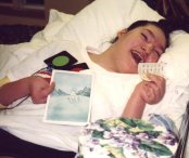
Amanda the reason CPN was
started
CPN
Reunion 2003

CP
Research
What's happening Today with
Cerebral Palsy
Special
Olympics
What's happening with
Special Olympics in 2003

CP
& Education
What's happening with
Special Need and Education in 2003