|
|
Insect Wings
- Insect wings have rigid veins which support the wing in flight. The wing veins may look different in different insect groups, scientists tracked that all different insect wings are evolved from the same ancestor, i.e. wings had evolved only once in the insects history.

- Typical insect wing venation (modified Comstock-Needham System)
-
- In the insect history, the fossil records show that the early insect wing had 8 pairs of main veins. Each pair diverged from wing base into anterior convex and posterior concave sector (e.g. MA and MP). The two sectors often fused into one veinal stem near the wing base. In evolution insect wing is in most case modified in reduction of veins.
- Precoasta (PC) -- This vein is fused with costa in all extant insects, mostly unrecognisable.
- Costa (C) -- at the leading edge of the wing, strong and marginal, extends to the apex of the wing, it is unbranched.
- Subcosta (Sc) -- the second longitudinal vein, mainly the subcosta posterior sector (ScP). Sc is reduced or fused with R in most Hemiptera.
- Radius (R) -- the third vein, usually the strongest vein on the wing, with branches usually cover the largest area of wing apex. RP is often referred to as radial sector (Rs) and the end branches as R1-5.
- Media (M) -- the fourth longitudinal vein, MA and MP usually with 4 branches each. In some insect groups MA fused with R so only MP on the medial area. In this case the MP1-4 are often referred as M1-4.
- Cubitus (Cu) -- fifth longitudinal vein, CuA may branch to 4 or fewer veins. CuP is unbranched, lies near the claval fold and reach the wing posterior margin.
- Anal veins (A) -- veins behind the cubitus, AA and AP are usually separated by the anal fold. In Neoptera, AA is always fused with Cu or CuP. In the hind wings of most orthopteroid insects, there is a large anal area where anals branch several times to form a fan-like folded wing.
- Jugal (J) -- small veins in the jugal area, found only in Neoptera.
- The black pterostigma is carried near the wing tip, between RA1+2 and RA3+4.
- Cross-veins are transverse veins joining longitudinal veins. Their names are based on the position relative to longitudinal veins, e.g. r-m is the cross-vein between radius and media.
- The wing veins of different insect groups are listed below. More information about insect flight in this page.
- In the insect history, the fossil records show that the early insect wing had 8 pairs of main veins. Each pair diverged from wing base into anterior convex and posterior concave sector (e.g. MA and MP). The two sectors often fused into one veinal stem near the wing base. In evolution insect wing is in most case modified in reduction of veins.
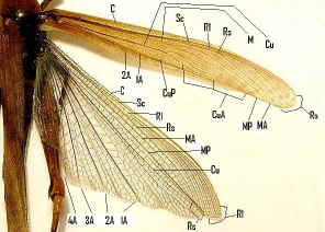
Damselfly and Dragonfly's Wings
- The Damselfly and Dragonfly both have two pairs of wings which are about
equal in size and shape. They are clean in colour.


- Wings of Australian Flatwing Damselfly Wings of male Australian Tiger Dragonfly
- There are five (R+M counted as 1) main vein stems on dragonfly and damselfly wings;
- Costa (C) -- at the leading edge of the wing, strong and marginal, extends to the apex of the wing.
- Subcosta (Sc) -- second longitudinal vein, it is unbranched, joins C at nodus.
- Radius and Media (R+M) -- third and fourth longitudinal vein, the strongest vein on the wing, with branches, R1-R4, reach the wing margin, the media anterior (MA) are also reach the wing margin. IR2 and IR3 are intercalary veins behind R2 and R3 respectively.
- Cubitus (Cu) -- fifth longitudinal vein, cubitus posterior (CuP) is unbranched and reach the wing margin.
- Anal veins (1A) -- unbranched veins behind the cubitus.
- A nodus is formed where the second main vein meets the leading edge of the wing. The black pterostigma is carried near the wing tip.


- The main veins and the crossveins form the wing venation pattern. The venation patterns are different in different species. There may be very numerous crossveins or rather few. The Australian Flatwing Damselfly's wings are one of the few veins patterns. The venation pattern is useful for species identification.
Cockroach's wings
- Cockroach's forewing are also known as tegmen, more or less sclerotised. It is used in flight as well as protection of membranous hind wings.

- Wing veins of Austral Ellipsidion Cockroach
- Costa (C) -- at the leading edge of the wing.
- Subcosta (Sc) -- second longitudinal vein, it is relatively short.
- Radius (R) -- third longitudinal vein, with many pectinate branches.
- Media (M) -- fourth longitudinal vein, reach the wing margin.
- Cubitus anterior (CuA) -- fifth longitudinal vein, with dichotomous branches occupy large part of tegmen.
- Cubitus posterior (CuP) is unbranched, curved and reach the wing margin.
- Anal veins (A) -- veins behind the cubitus.
- The veins of hind wing are about the same as front wing but with large anal lobe folded at rest between CuP and 1A. The anal lobe usually folded in a fan-like manner.
- Subcosta (Sc) -- second longitudinal vein, it is relatively short.

Grasshopper's Wings
- Grasshopper forewings are tough opaque tegmina, narrow and covering the hind wings
and abdomen at rest. Hind wings are board membranous and
folded in fan-like manner.
- Wing veins of Giant Grasshopper
- Costa (C) -- at the leading marginal of the forewing and hind wing, unbranched.
- Subcosta (Sc) -- second longitudinal vein, unbranched.
- Radius (R) -- third longitudinal vein, branched to Rs in forewing and hind wing.
- Media anterior (MA) -- fourth longitudinal vein, branched in basal part as Media posterior (MP).
- Cubitus (Cu) -- fifth longitudinal vein, on forewing and hind wing dividing near the wing base into branched CuA, and unbranched CuP.
- Anal veins (A) -- veins behind the cubitus, unbranched, two in forewing, many in hind wing.

Stick Insect's Wings
-
Stick insect forewings are tough opaque tegmina, short and covering only the
base part of the hind wings at rest. Hind wings from costa to Cubitus are
tough and opaque like the forewings. The large anal area are membranous and
folded in fan-like manner. There are no or very few branching in Stick Insect
wing veins.


- Wing veins of Children's Stick Insect Wing veins of male Goliath Stick Insect
- Costa (C) -- at the leading marginal of the hind wing, unbranched, absent in forewing.
- Subcosta (Sc) -- second longitudinal vein, unbranched.
- Radius (R) -- third longitudinal vein, branched to Rs in hind wing, unbranched in forewing.
- Media anterior (MA) -- fourth longitudinal vein, branched in basal part as Media posterior (MP).
- Cubitus (Cu) -- fifth longitudinal vein, unbranched.
- Anal veins (A) -- veins behind the cubitus, unbranched, two in forewing, many in hind wing 1A-7A in one group and the rest in another group.


Cicada's Wings
-
Both forewings and hindwings of Cicada are membranous, most species are
glass-like although some are opaque. Cicada is not a good flier and most fly
only a few seconds. When fly, forewing and hind wing are hooked together by a
grooved coupling along the hind wing costa and forewing margin. Most species
have a basic venation as shown in the following picture.

- Wing veins of Cicada
- Costa (C) -- at the leading wing marginal, in forewing extends to the node and lies close to Sc+R.
- Subcosta + Radius (Sc+R) -- in forewing Sc and R fused together to the node. Radial sector (Rs) arises near the node and unbranches.
- Radius anterior (RA)
- Radius posterior (RP)
- Media (M) -- branches to M1 to M4.
- Cubitus anterior (CuA) -- branches to CuA1 and CuA2.
- Cubitus posterior (CuP) -- unbranches.
- Anal veins (A) -- veins behind the cubitus, 1A and 2A fused in the forewing, CuP and 2A are folded.
- Also notice there are the ambient veins and peripheral membranes on the margin of both wings.

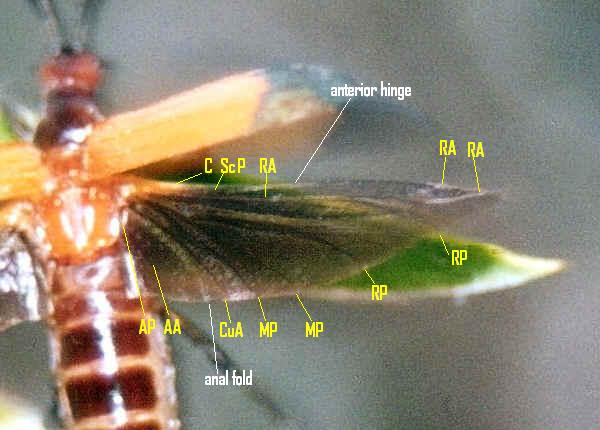
Beetle's Wings
- Beetle's wing cover, also know as elytra, is the modified forewing. It is
rigid, covering over the abdomen when at rest. When flight, it is held out at
an angle with little movement, very little contributing to the aerodynamic.
- Beetle's functional wings are the hind wings. The hind wing is longer than the elytra, folded longitudinally and transversely under the elytra. The wing is rotated forwards on its base into flight position. This action spread the wing and unfolded longitudinally and transversely. There is the spring mechanism in the wing structure, sometimes with the help of abdomen movement, to keep the wing in folded position.

- Wing veins of Beetle
- Beetle wing venation is reduced and modified due to the folding structure.
- Costa (C), Subcosta posterior (ScP) -- at the leading wing marginal, fused for most of the length.
- Radius anterior (RA) - divided into two branches beyond the middle of the wing.
- Radius posterior (RP) - basal connection is lost.
- Media posterior (MP) -- branches, long and strong vein.
- Cubitus anterior (CuA)
- Anal veins (AA, AP) -- veins behind the cubitus, separated by anal fold.
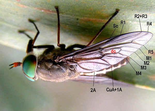
Fly's Wings
-
Fly has only one pair of functional wings. The hind wings reduced to small
club-like structure known as halteres. The halteres vibrate rapidly during
flight, act as gyroscopic sense organs of balance.


- Wing veins of Tipulidae Wing veins of Tabanoidae


- Wing veins of Asilidae Wing veins of Bombyliidae


- Wing veins of Syrphidae Wing veins of Tachinidae
- Costa (C) -- not found in Diptera.
- Subcosta (Sc) -- became the leading wing vein, unbranched.
- Radius (R) -- branched to R1-R5.
- Media (M) -- branched to M1-M4.
- Cubitus (CuA) -- unbranched, CuP is reduced in Diptera. Some species CuA and 1A are separated, some species meets when reaching the wing margin, some species fused.
- Anal veins (A) -- only two anal veins 1A and 2A are present, 2A is not distinctive in some species.
- Discal Cell (dc) -- well define in most species.


Butterfly's Wings
-
Butterfly has two pairs of membranous wings, clothed with with two layers of
'scales'.


- Wing veins of PAPILIONIDAE Wing veins of PIERIDAE


- Wing veins of NYMPHALIDAE Wing veins of LYCAENIDAE
- Costa (C) -- not found in Butterflies.
- Subcosta + Radius 1 (Sc+R1) -- at the leading wing marginal, fused or very close for most of the length, in hind wing fused and well developed in the humeral area, subcosta never branches in butterfly.
- Radius (R2-R5) - radius divides into branches beyond the middle of the wing up to five branches in Papilionidae. On forewing, the last R is stalked in all butterflies except Hesperiidae is separated.
- Radius sector (Rs) - in hind wing.
- Media (M1-M3) - the basal section has been lost.
- Cubitus anterior (CuA1-CuA2) - CuP section has been lost.
- Anal veins (A, 1A+2A, 3A) - either one vein A, or two veins 1A+2A, 3A.
- Humeral vein - Notice that the hind wing of most butterflies (except Lycaenidae) has the humeral vein. There is the enlargement of the humeral area of the hind wing which is overlapped with the fore wing. The humeral vein strengthened the hind wing overlapped area so that the two wings coupling better.


Hymenoptera Wing
- The Hymenoptera adults, include saw flies, wasps, bees and ants (except the working ants), has two pair of membranous wings


- Wing veins of Sawfly Wing veins of wasp

- Wing veins of Ant
-
- Costa (C) -- not found in Hymenoptera.
- Subcosta (Sc) -- unbranched.
- Radius (R) -- branched to R1-R5.
- Media (M) -- M is unbranches, in forewing M is fused with Rs for part of its length.
- Cubitus (CuA) -- unbranched, CuP is absent in Hymenoptera.
- Anal veins (A) -- only two anal veins 1A and 2A are present, 2A is not distinctive in some species.
- Wing-coupling - Row of hooks on the leading edge of hind wing engage the hind margin of the forewing, strongly couple the wings in flight.
- Line of wing folding - Some species, including Vespidae, the forewing are longitudinally folded along the 'line of wing folding' at rest.
- Pterostigma is present for some species.
- Costa (C) -- not found in Hymenoptera.


- Reference:
- 1. Insects of Australia, CSIRO, Division of Entomology, Melbourne University Press, 2nd Edition 1991, pp 15, 295, 321, 370, 432, 725, 819, 921.
- 2. The Australian dragonflies, J.A.L.Watson, G. Theischinger, H.M. Abbey, CSIRO 1991, pp 58.
- 3. Dragonflies, P.L. Miller, Cambridge University Press, 1987, pp 24.
- 4. External Anatomy - WINGS - John R. Meyer, Department of Entomology, NC State University, 19 September 2002.
- 5. A Guide to Australian Grasshoppers and Locusts - D.C.F.Rentz, R.C.Lewis, Y.N.Su and M.S.Upton, Natural History Publications (Borneo), 2003, pp 26.
- 6. Australian Cicadas - M.S. Moulds, Australian Museum, UNSW Press, 1990, pp 15.
- 7. The Complete Field Guide to Butterflies of Australia - Michael F. Braby, Australian National University, CSIRO PUBLISHING, 2004, pp 6.
- 8. Animal
Flight - Department of Zoology,
Back to Top
